© UncutMed — Medical knowledge, precise, truthful, unfiltered. The current article was last modified on October 18, 2026. This article was clinically authored and reviewed under international evidence-based standards. Always consult your personal healthcare physician before modifying any medical therapy.
Medical Disclaimer: This article is for informational and educational purposes only. It does not constitute medical advice, formal diagnosis, or treatment recommendations. Always consult a qualified healthcare professional regarding any metabolic condition or therapeutic plan.
1. Pulmonary Embolism Clinical Definition
Section Summary:
Pulmonary embolism is an acute, life-threatening cardiovascular emergency caused by the mechanical obstruction of the pulmonary arterial bed, predominantly by a dislodged venous thrombus. This occlusion elevates right ventricular afterload, disrupts pulmonary gas exchange, and compromises systemic perfusion. Recognizing its pathophysiological mechanism, immediate clinical presentation, and hemodynamic impact is vital for rapid risk stratification, early therapeutic intervention, and the prevention of catastrophic right ventricular failure and sudden cardiovascular collapse.
Pulmonary Embolism (PE) is defined as the acute occlusion of one or more branches of the pulmonary arterial bed by endogenous or exogenous material originating from a distant vascular site. In more than 90% of clinical cases, the embolic nidus consists of a dislodged thrombus arising from the deep venous system of the lower extremities or pelvis—a pathological spectrum collectively designated as Venous Thromboembolism (VTE).
From an anatomical and hemodynamic perspective, pulmonary embolism represents a critical breakdown of both the pulmonary and systemic circulatory systems. The pulmonary circulation is intrinsically a low-resistance, high-capacitance vascular circuit engineered to accommodate the entire right ventricular stroke volume at low pressures (mean pulmonary arterial pressure ≤ 20 mmHg at rest). When an embolus lodges in the main pulmonary trunk, the right or left main pulmonary arteries, or downstream lobar, segmental, and subsegmental branches, it suddenly transforms this low-resistance circuit into a high-resistance vascular bed.
Clinical classification categorizes Pulmonary Embolism based on anatomical extent and hemodynamic consequence:
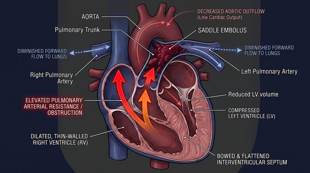
- Saddle Pulmonary Embolism: A massive clot straddling the bifurcation of the main pulmonary artery trunk, extending simultaneously into both the right and left main pulmonary arteries. It presents an imminent risk of total circulatory arrest.
- Lobar and Segmental Embolism: Occlusion localized to intermediate-caliber branches, frequently producing distinct ventilation-perfusion mismatch and localized pulmonary infarction.
- Subsegmental Embolism: Obstruction confined to peripheral terminal vessels, which may spare overall right ventricular hemodynamics but can provoke acute pleuritic irritation and localized alveolar hemorrhage.
Functionally, modern international consensus guidelines (European Society of Cardiology [ESC] and the American Heart Association [AHA]) classify Pulmonary Embolism (PE) not merely by anatomical clot size, but by early mortality risk driven by right ventricular (RV) failure and hemodynamic instability:
- High-Risk (Massive) Pulmonary Embolism: Sustained systemic hypotension (systolic blood pressure < 90 mmHg or a drop of ≥ 40 mmHg for longer than 15 minutes not caused by new-onset arrhythmia, hypovolemia, or sepsis), obstructive shock, or cardiac arrest.
- Intermediate-Risk (Submassive) Pulmonary Embolism: Hemodynamically stable (systolic blood pressure ≥ 90 mmHg), but presenting with evidence of right ventricular dysfunction on imaging (echocardiography or CT) and/or myocardial injury confirmed by elevated cardiac biomarkers (Troponin I/T or NT-proBNP). This is sub-stratified into Intermediate-High (both RV dysfunction and elevated biomarkers) and Intermediate-Low (either RV dysfunction or elevated biomarkers, but not both).
- Low-Risk Pulmonary Embolism: Hemodynamically stable patients without evidence of right ventricular overload, myocardial necrosis, or significant clinical comorbidities.
2. Pulmonary Embolism Causes and Etiology
Section Summary:
Pulmonary embolism primarily arises from detached deep vein thrombi originating in the proximal lower extremities or pelvis. Less common non-thrombotic etiologies include fat emboli following long-bone orthopedic fractures, amniotic fluid emboli during labor, air embolization from central vascular access, and septic emboli from right-sided infective endocarditis. All cause mechanical vascular occlusion, triggering an acute inflammatory cascade and downstream pulmonary endothelial ischemia.
The vast majority of pulmonary emboli originate as a downstream consequence of Deep Vein Thrombosis (DVT). However, a wide spectrum of non-thrombotic materials can embolize into the pulmonary arterial tree, each exhibiting distinct pathophysiological and clinical signatures.
Thromboembolic Etiology
- Proximal Lower Extremity Deep Vein Thrombosis: Responsible for approximately 75% to 80% of clinically significant Pulmonary Embolisms. Thrombi formed within the popliteal, femoral, and external iliac veins possess substantial volume and a high propensity for spontaneous dislodgement during muscle contraction or sudden changes in intrathoracic pressure. Most of these types of emboli form in the gastrocnemius anatomical region of the legs.
- Pelvic and Deep Abdominal Veins: Thrombi developing within the internal iliac, prostatic, uterine, and ovarian venous plexuses, frequently triggered by pelvic surgery, peripartum states, or pelvic malignancies.
- Distal (Calf) Deep Vein Thrombosis: Thrombi restricted to the posterior tibial, peroneal, and anterior tibial veins. While isolated calf thrombi embolize less frequently (< 10%), approximately 20% to 30% propagate proximally into the popliteal fossa if left untreated, subsequently becoming major embolic threats.
- Upper Extremity DVT (UEDVT): Accounts for 5% to 10% of Pulmonary Embolism (PE) events. These are predominantly iatrogenic, precipitated by indwelling central venous catheters (CVCs), peripherally inserted central catheters (PICCs), transvenous cardiac pacemaker or defibrillator leads, or anatomical vascular compression at the thoracic outlet (Paget-Schroetter syndrome).
Non-Thrombotic Embolic Etiologies
- Fat Embolism Syndrome (FES): Typically occurs 24 to 72 hours following traumatic fractures of long bones (especially the femur and tibia) or major orthopedic pelvic reconstruction. Bone marrow disruption releases neutral fat globules into ruptured intramedullary sinusoids. In addition to mechanical capillary occlusion, free fatty acids generate extensive endothelial cytotoxicity, causing systemic petechiae, acute respiratory distress, and encephalopathy.
- Amniotic Fluid Embolism (AFE): A rare, catastrophic obstetric emergency occurring during labor or the immediate postpartum period. Breaches in the maternal-fetal barrier allow fetal squamous cells, lanugo, and procoagulant amniotic fluid to enter maternal pelvic veins. This induces a devastating, biphasic reaction: profound transient pulmonary vasoconstriction with acute right heart failure, followed by severe Disseminated Intravascular Coagulation (DIC) and massive hemorrhage.
- Air / Gas Embolism: Iatrogenic introduction of air into the venous system through accidental infusion during central line placement, line removal, laparoscopic insufflation, or cardiothoracic surgery. A bolus of 100 to 200 mL of air can form an intractable physical “air lock” within the right ventricular outflow tract (RVOT), completely halting forward pulmonary perfusion.
- Septic Embolism: Clots laden with bacterial or fungal microcolonies that fragment from infected intravascular foci, classic in tricuspid valve infective endocarditis (common in intravenous drug users), infected central catheters, or septic thrombophlebitis of the internal jugular vein (Lemierre’s syndrome). These manifest as recurrent, wedge-shaped peripheral infarcts that frequently cavitate into pulmonary abscesses.
- Tumor Embolism: Hematogenous dissemination of microscopic or macroscopic tumor clusters (e.g., renal cell carcinoma growing into the inferior vena cava, hepatocellular carcinoma, or gastric adenocarcinoma), leading to widespread microvascular occlusion and subacute pulmonary hypertension.
- Foreign Body and Cement Embolism: Methylmethacrylate bone cement leaking into the paravertebral venous system during percutaneous vertebroplasty or kyphoplasty, resulting in physical polymer cast occlusion of pulmonary arterioles.
3. Pulmonary Embolism Pathophysiology (Extensive Analysis) – The Hemodynamic and Respiratory Cascade
Section Summary:
The pathophysiology of pulmonary embolism centers on acute mechanical vascular obstruction and reactive humoral vasoconstriction, dramatically elevating pulmonary vascular resistance. This acute increase in right ventricular afterload causes right ventricular dilation, tricuspid regurgitation, and leftward interventricular septal bowing. Consequent left ventricular underfilling precipitates cardiogenic shock, while ventilation-perfusion mismatch, intrapulmonary shunting, and alveolar dead-space enlargement produce profound refractory arterial hypoxemia and cellular hypoxia.

The clinical consequences of pulmonary embolism are determined by the interplay between mechanical obstruction of the pulmonary vascular tree, neurohumoral reflex vasoconstriction, and the patient’s baseline cardiopulmonary functional reserve.
1. Hemodynamic Derangements: The Right Ventricular Spiral of Death
The unconditioned right ventricle is a thin-walled, compliant chamber designed solely to generate low systolic pressures (20–25 mmHg). It is physiologically unequipped to match sudden, dramatic surges in afterload:
- Vascular Obstruction and Vasoconstrictive Humoral Mediators: As thrombi lodge within the pulmonary arterial circulation, physical occlusion of the vascular lumen is compounded by intense, active vasoconstriction. Activated platelets adhering to the thrombus release vasoactive mediators, including Thromboxane A2 (TXA2) and Serotonin (5-HT). Concurrently, dysfunctional pulmonary endothelial cells discharge Endothelin-1 and suppress endothelial nitric oxide synthase (eNOS) activity. This biochemical surge amplifies downstream vasoconstriction, causing Pulmonary Vascular Resistance (PVR) to skyrocket.
- Acute Right Ventricular Afterload Mismatch and Dilation: If the mechanical and vasoconstrictive obstruction exceeds 30% to 50% of the total pulmonary arterial cross-sectional area, pulmonary arterial pressure climbs sharply. Because the normal, non-hypertrophied Right Ventricle (RV) cannot acutely generate a mean pulmonary artery pressure much higher than 40 mmHg, it responds by dilating.
- Right Ventricular Wall Stress and Myocardial Ischemia: In accordance with Laplace’s Law (σ = P*r/2*h, where wall stress “σ” is directly proportional to intraventricular pressure “P” and internal radius “r” and inversely proportional to wall thickness “h”), acute Right Ventricular dilation markedly elevates wall tension. Increased wall tension sharply drives up Right Ventricular myocardial oxygen demand (MVO2).
- Impairment of Right Ventricular Coronary Perfusion: Unlike the left ventricle (which is perfused almost entirely during diastole), the right ventricle is normally perfused during both systole and diastole because systemic aortic pressures consistently exceed RV intracavitary pressures. In acute massive Pulmonary Embolism (PE), the combination of elevated RV intracavitary diastolic pressure and decreased systemic arterial pressure compresses the right coronary artery (RCA) microvasculature. Transmural RV perfusion pressure drops, precipitating acute Right Ventricle subendocardial ischemia, cardiogenic myocyte necrosis, and the release of cardiac troponins.
- Interventricular Interdependence and Left Ventricular Collapse: The dilated, failing right ventricle shifts the interventricular septum (an anatomical structure that separates the right ventricle from the left ventricle of the heart) toward the left ventricle (termed paradoxical septal bowing or the reverse Bernheim effect). This septal displacement encroaches upon the left ventricular cavity, severely distorting its geometry, reducing Left Ventricle (LV) compliance, and impairing LV diastolic filling (decreased LV preload).
- Cardiogenic Shock and Circulatory Arrest: Decreased Left Ventricle preload leads directly to a fall in stroke volume and systemic cardiac output. Systemic hypotension depresses aortic root perfusion pressure, further starving the right coronary artery of blood. This establishes a lethal positive feedback loop—the “RV spiral of death”—terminating in profound obstructive cardiogenic shock, electromechanical dissociation (pulseless electrical activity), and fatal cardiovascular collapse.
2. Respiratory Pathophysiology and Impairment of Gas Exchange
Pulmonary embolism compromises respiration through three primary mechanisms:
- Alveolar Dead-Space Ventilation (V/Q tends to go close to ∞): Occlusion of pulmonary arterial branches abruptly ceases perfusion to ventilated lung units. These alveoli continue to be ventilated without participating in gas exchange, producing a sharp expansion of anatomical and physiological dead space. This wasted ventilation impairs the efficient excretion of carbon dioxide, requiring compensatory hyperventilation.
- Ventilation-Perfusion Mismatch (V/Q < 1) and Intrapulmonary Shunting: Total cardiac output is diverted away from obstructed zones into remaining patent pulmonary vessels. These hyperperfused vascular zones are overwhelmed by blood flow relative to their ventilatory capacity, generating areas of low ventilation-to-perfusion ratios (V/Q << 1). Furthermore, downstream alveolar surfactant depletion causes atelectasis within 24 to 48 hours, creating true intrapulmonary shunts where blood flows through completely unventilated alveoli. This causes a widening of the Alveolar-arterial oxygen gradient and hypoxemia that does not fully correct with supplemental oxygen alone.
- Reflex Tachypnea and Hyperventilation: Mechanical stretch and ischemia stimulate pulmonary neuroreceptors (intrapulmonary J-receptors and irritant receptors), triggering a vagally mediated reflex drive to hyperventilate. This reflex produces marked tachypnea, hypocapnia, and an acute respiratory alkalosis.
4. Pulmonary Embolism Symptoms
Section Summary:
The clinical symptoms of pulmonary embolism vary from subtle dyspnea to sudden cardiovascular collapse, depending on clot burden and underlying cardiopulmonary reserve. Common manifestations include sudden-onset dyspnea from ventilation-perfusion mismatch, pleuritic chest pain from pleural irritation or peripheral pulmonary infarction, hemoptysis due to alveolar hemorrhage, and presyncope or syncope resulting from acute right ventricular outflow tract obstruction and transient systemic cerebral hypoperfusion.
The clinical presentation of Pulmonary Embolism (PE) varies widely, earning its reputation as “the great masquerader.” Symptoms can range from isolated, indolent breathlessness to sudden cardiovascular collapse.
| Clinical Symptom | Prevalence (%) | Detailed Pathophysiological Mechanism |
| Dyspnea | 75% – 85% | Abrupt-onset breathlessness driven by acute V/Q mismatch, arterial hypoxemia, dead-space expansion, and vagally mediated stimulation of pulmonary J-receptors and irritant stretch receptors. |
| Pleuritic Chest Pain | 65% – 75% | Typically caused by smaller, peripheral emboli occluding subsegmental branches. The resulting localized pulmonary infarction causes focal ischemia and secondary inflammation of the richly innervated parietal pleura, causing sharp pain aggravated by inspiration. |
| Retrosternal Anginal Chest Pain | 10% – 15% | Characteristic of central, massive Pulmonary Embolism. Unlike peripheral pleuritic pain, this is a dull, heavy, crushing substernal ache caused by acute right ventricular ischemia, driven by excessive wall tension and critically diminished right coronary artery perfusion. |
| Cough | 35% – 40% | Triggered by localized inflammatory irritation of the tracheobronchial tree, alveolar atelectasis, and neurochemical activation of sensory fibers by locally released autacoids (histamine, prostaglandins). |
| Hemoptysis | 10% – 15% | Blood-tinged or overtly bloody sputum resulting from distal pulmonary infarction. Ischemic necrosis of the alveolar-capillary membrane leads to intra-alveolar hemorrhage that spills into the conducting airways. |
| Syncope / Presyncope | 10% – 12% | Transient global cerebral hypoperfusion caused by a sudden, critical drop in cardiac output. This indicates a large clot burden (saddle or main pulmonary artery occlusion) that mechanically limits right ventricular outflow and precipitates systemic hemodynamic collapse. |
| Unilateral Lower Limb Pain / Swelling | 25% – 30% | Direct clinical manifestation of the underlying deep vein thrombosis. Mechanical venous obstruction and secondary perivenous inflammation elevate hydrostatic pressure, causing localized edema and pain. |
5. Pulmonary Embolism Clinical Signs
Section Summary:
Physical examination in pulmonary embolism reflects acute sympathetic stimulation, pulmonary hypertension, and right-sided circulatory compromise. Tachypnea and resting sinus tachycardia are the most frequent objective physical findings. Severe hemodynamic impairment manifests through jugular venous distention with prominent waves, an accentuated pulmonic heart sound (P2), a right ventricular S3 gallop, hypotension, cool, clammy extremities, and asymmetric unilateral lower extremity edema indicating concurrent deep vein thrombosis.

The physical examination in acute Pulmonary Embolism (PE) reflects the body’s compensatory sympathetic neurohumoral response to maintain perfusion, alongside direct evidence of acute right heart overload.
- Tachypnea (Respiratory Rate > 20 breaths/min): Present in over 70% of patients. It represents the primary compensatory mechanism for expanding alveolar dead space and hypercapnic drive, amplified by neurochemical stimulation of pulmonary vagal afferents.
- Tachycardia (Heart Rate > 100 beats/min): Driven by an intense endogenous surge of catecholamines (epinephrine and norepinephrine) attempting to sustain cardiac output (Cardiac Output = Heart Rate * Stroke volume) in the face of a falling stroke volume.
- Hypoxemia and Cyanosis: Decreased peripheral oxygen saturation (SpO2 < 90% on ambient air). Central cyanosis may develop in massive Pulmonary Embolism (PE) due to severe intrapulmonary shunting, low mixed venous oxygen saturation (SvO2), or a patent foramen ovale (PFO) opening right-to-left under high right atrial pressures.
- Jugular Venous Distension (JVD): Elevated jugular venous pressure with prominent “a” waves (from forceful right atrial contraction against high RV end-diastolic pressure) and “v” waves (from functional tricuspid regurgitation). A positive Kussmaul sign (paradoxical rise in JVP during inspiration) may occasionally be observed.
- Accentuated Pulmonic Component of S2 (Loud P2): Auscultated at the left second intercostal space. Caused by high back pressure in the main pulmonary artery snapping the pulmonic valve leaflets closed abruptly at end-systole.
- Tricuspid Regurgitation Murmur & Right Ventricular S3 Gallop: A holosystolic murmur heard best at the left lower sternal border that intensifies during inspiration (Carvallo’s sign), caused by severe annular dilation of the tricuspid valve. An RV third heart sound reflects volume overload and non-compliant diastolic filling of the failing right ventricle.
- Left Parasternal Lift (Right Ventricular Heave): A palpable systolic lift along the left sternal margin, produced by the hyperdynamic, dilated right ventricle contracting directly against the anterior chest wall.
- Systemic Hypotension and Signs of Shock: Systolic blood pressure < 90 mmHg, oliguria, altered mental status, and cool, pale, clammy extremities with prolonged capillary refill (> 3 seconds). These findings indicate critical cardiac decompensation and obstructive shock.
- Low-Grade Fever: Body temperature ranging between 37.5 and 38.5 degrees Celsius (or 99.5 to 101.3 degrees Fahrenheit)
6. Pulmonary Embolism Risk Factors and Stratification (Virchow’s Triad)
Section Summary:
Risk factors for pulmonary embolism align with Virchow’s triad: endothelial injury, venous stasis, and hypercoagulability. Major provoking factors include major orthopedic or abdominal surgery, prolonged immobilization, polytrauma, active malignancy, and pregnancy or estrogen-containing hormonal therapies. Inherited thrombophilias such as Factor V Leiden and prothrombin mutations, alongside acquired conditions like antiphospholipid syndrome and advanced systemic atherosclerosis, significantly amplify baseline thromboembolic susceptibility across susceptible populations.
The development of pulmonary embolism is fundamentally anchored in Virchow’s Triad—the three converging pathological pillars of intravascular thrombosis:
- Vascular Endothelial Injury: Exposing subendothelial collagen and tissue factor, triggering the extrinsic coagulation cascade.
- Venous Stasis: Disrupting laminar blood flow, preventing the clearance of activated clotting factors, and facilitating local thrombin accumulation.
- Hypercoagulability: Genetic or acquired prothrombotic imbalances that favor coagulation over natural anticoagulant and fibrinolytic pathways.
Vascular Endothelial Injury can occur through many incidents:
- Injury or surgery
- Venipuncture
- Chemical irritation
- Heart valve disease or heart valve replacement
- Atherosclerosis
- Permanent catheter
Venous stasis is a process that can happen in various ways:
- Atrial Fibrillation
- Left Ventricle malfunction
- Immobility or paralysis
- Venous insufficiency or varicose veins
- Venous obstruction due to a neoplasm, excess weight (obesity), or pregnancy
Hypercoagulability is a state that can be caused by a variety of reasons:
- Malignancy
- Pregnancy and perinatal period
- Estrogen therapy
- Trauma and surgery in the lower limbs, the hips, the abdomen, or the pelvis
- Inflammatory bowel disease
- Nephrotic syndrome
- Sepsis
- Thrombophilia
Clinical practice classifies risk factors for Pulmonary Embolism (PE) into strong, moderate, and weak contributors based on their odds ratio (OR) for triggering an acute event:
Pulmonary Embolism Strong Risk Factors
Strong risk factors for Pulmonary Embolism have an Odds Ratio > 10:
- Major orthopedic surgery (total hip arthroplasty, total knee replacement)
- Major trauma, polytrauma, or extensive spinal cord injury with paraplegia
- Fractures of the lower limbs (femur, tibia, fibula)
- Hospitalization for acute heart failure or atrial fibrillation within the previous 3 months
- Previous documented venous thromboembolism (Deep Vein Thrombosis or Pulmonary Embolism)
- Myocardial Infarction within the previous 3 months
- Spinal Cord injury
Pulmonary Embolism Moderate Risk Factors
Moderate risk factors for Pulmonary Embolism have an Odds Ratio of 2 – 9:
- Chemotherapy, biological anti-neoplastic agents, or checkpoint inhibitors
- Central venous lines, PICC lines, or transvenous pacemaker leads
- Arthroscopic knee surgery
- Autoimmune inflammatory disorders (Systemic Lupus Erythematosus, Rheumatoid Arthritis, IBD: Crohn’s/Ulcerative Colitis)
- Inherited thrombophilias (Factor V Leiden, Prothrombin gene mutation G20210A, Antithrombin deficiency, Protein C or S deficiency)
- Acquired thrombophilias (Antiphospholipid Syndrome: lupus anticoagulant, anti-cardiolipin antibodies, anti-β2-glycoprotein I)
- Pregnancy and early postpartum period (highest risk within the first 6 weeks post-delivery)
- Active malignancy (especially adenocarcinomas: pancreatic, gastric, lung, ovarian, and brain neoplasms)
- Contraceptive pills
- Acute systemic infections (pneumonia, sepsis, severe COVID-19 infection)
- Neurological disorders that cause paralysis
- Blood transfusion and the use of erythropoietin
- Chronic Heart Failure or Respiratory Failure
Pulmonary Embolism Weak Risk Factors
Weak risk factors for Pulmonary Embolism have an Odds Ratio < 2:
- Bed rest or physical immobility > 3 days
- Prolonged travel / long-haul air flights (> 4 – 6 hours continued sitting)
- Advanced age (> 65 years, risk doubles each subsequent decade)
- Excess weight (Obesity)
- Surgery with laparoscopy
- Varicose veins and superficial venous insufficiency
- Essential hypertension, diabetes mellitus, and systemic atherosclerosis
Provoked vs Unprovoked Stratification
- Provoked Pulmonary Embolism: Occurs secondary to a recognized, temporary major clinical risk factor (e.g., major surgery under general anesthesia > 30 minutes, trauma, or prolonged immobilization). Once the provoking trigger resolves, the recurrence rate is low (~ 1 – 3 per year).
- Unprovoked (Idiopathic) PE: Occurs in the complete absence of identifiable temporary risk factors, or in the presence of persistent, irreversible drivers (e.g., metastatic cancer, persistent antiphospholipid syndrome). These patients carry a high long-term recurrence risk (~ 8% – 10% at 1 year, and > 30% at 5 years), often requiring extended or indefinite anticoagulation.
7. Pulmonary Embolism Diagnosis
Section Summary:
Diagnosing pulmonary embolism requires structured pre-test probability assessment using the Wells or Geneva score combined with age-adjusted D-dimer testing to exclude low-risk cases. Computed Tomography Pulmonary Angiography represents the definitive imaging gold standard, while V/Q scintigraphy serves contrast-allergic patients. Electrocardiography reveals right ventricular strain patterns, chest radiographs rule out alternative thoracic pathologies, and elevated troponins or natriuretic peptides identify acute subclinical myocardial injury.

Diagnosing pulmonary embolism requires a systematic, validated approach. Because presenting symptoms are non-specific, relying on clinical impression alone leads to missed diagnoses and unnecessary, expensive imaging.
1. Clinical Pre-Test Probability Assessment
Before ordering laboratory testing or advanced imaging, clinicians must quantify the patient’s pre-test probability using a validated scoring tool: the Wells Score for Pulmonary Embolism or the Revised Geneva Score.
The Wells Criteria (Wells score) for Pulmonary Embolism (PE)
| Clinical Variable | Assigned Points |
| Clinical signs and symptoms of Deep Vein Thrombosis (minimum of leg swelling and pain with palpation) | + 3.0 |
| Alternative diagnosis is less likely than pulmonary embolism | + 3.0 |
| Heart rate > 100 beats per minute | + 1.5 |
| Immobilization (≥ 3 consecutive days) or surgery within the previous 4 weeks | + 1.5 |
| Previous objectively diagnosed Pulmonary Embolism or Deep Vein Thrombosis | + 1.5 |
| Hemoptysis | + 1.0 |
| Malignancy (treatment ongoing, within 6 months, or receiving palliative care) | + 1.0 |
Three-Tier model:
- Low Probability: 0 – 1 point (incidence of PE ~ 10%)
- Intermediate Probability: 2 – 6 points (incidence of PE ~ 30%)
- High Probability: ≥ 7 points (incidence of PE ~ 65%)
Two-Tier model:
- Pulmonary Embolism Unlikely: ≤ 4 points (proceed to D-dimer testing)
- Pulmonary Embolism Likely: ≥ 5 points (bypass D-dimer testing, proceed directly to CTPA imaging)
The Geneva criteria (Geneva score) for Pulmonary Embolism (PE)
| Clinical Variable | Assigned points |
|---|---|
| Heart Rate > 95 beats per minute | + 5.0 |
| Clinical signs and symptoms of Deep Vein Thrombosis (minimum of leg swelling and pain with palpation) | + 4.0 |
| Heart Rate 75 – 94 beats per minute | + 3.0 |
| Single-sided pain of the lower limb | + 3.0 |
| Previous objectively diagnosed Pulmonary Embolism or Deep Vein Thrombosis | + 3.0 |
| Surgery or bone fracture (< 4 weeks) | + 2.0 |
| Hemoptysis | + 2.0 |
| Cancer | + 2.0 |
| Age > 65 | + 1.0 |
Likelihood for Pulmonary Embolism (PE):
- Low probability: 0 – 3 points
- Intermediate probability: 4 – 10 points
- High Probability: ≥ 11 points
The Pulmonary Embolism Rule-Out Criteria (PERC)
In patients categorized as low risk, the PERC rule can be applied. If the patient meets all eight of the following criteria, the probability of Pulmonary Embolism (PE) is < 1.8%, and all further diagnostic workups – including D-dimer – can safely be omitted:
- Age < 50 years
- Heart Rate < 100 beats per minute
- Oxygen saturation SqO2 ≥ 95% on room air
- No prior history of Deep Vein Thrombosis or Pulmonary Embolism
- No recent trauma or surgery requiring hospitalization within past 4 weeks
- Hemoptysis
- No exogenous estrogen use
- No unilateral lower extremity swelling
2. General and Advanced Laboratory Investigations
D-Dimer testing:
- A degradation product of cross-linked fibrin produced during endogenous fibrinolysis.
- High Sensitivity (> 95%) but Low Specificity: Highly effective for ruling out Pulmonary Embolism, but elevated values occur in cancer, pregnancy, infection, inflammation, advanced age, surgery, and renal failure.
- Standard Cutoff: < 500 μg/L (or 0.5 mg/L Fibrinogen Equivalent Units [FEU]), when the measurement is done by a high-sensitivity method (such as ELISA).
- D-Dimer specificity becomes lower as people age, and it can reach just 10% at 80 years of age. Age-Adjusted D-Dimer Cutoff (for patients > 50 years) is calculated as: Cutoff = Age * 10 μg/L. Applying this age-adjusted formula significantly increases diagnostic specificity in elderly patients without compromising negative predictive value.
- When the clinical probability for Pulmonary Embolism is high, D-Dimers are NOT assessed.
Complete Blood Count (CBC):
- May demonstrate moderate leukocytosis with a left shift driven by systemic inflammatory cytokine release. Polycythemia may reflect chronic hypoxemia, while severe anemia can lower the threshold for myocardial ischemia. Please read our blood test article for a better understanding.
Renal Function (Urea, Serum Creatinine, eGFR) and Electrolytes:
- Essential for screening before administering iodinated intravenous contrast for CTPA (Computed Tomography Pulmonary Angiography).
- Establishes baseline clearance to guide dosing for low-molecular-weight heparins (LMWH) and direct oral anticoagulants (DOACs), which rely on renal excretion.
- Hyponatremia (Serum Na < 135 mEq/L) in acute Pulmonary Embolism (PE) serves as a recognized surrogate for severe neurohormonal activation and an independent predictor of in-hospital mortality.
Cardiac Biomarkers:
- Cardiac Troponins (Troponin I and T): Reflect acute right ventricular subendocardial ischemia and micro-infarction. Elevated levels indicate intermediate-to-high-risk disease.
- Natriuretic Peptides (BNP and NT-proBNP): Released by ventricular myocytes in response to acute stretch and wall stress. Elevated levels indicate clinically meaningful right ventricular overload.
Arterial Blood Gas:
- Classic triad: Acute Arterial Hypoxemia (PaO2 < 80 mmHg), Hypocapnia (PaCO2 < 35 mmHg), and Acute Respiratory Alkalosis (pH > 7.45) secondary to hyperventilation.
- An elevated alveolar-arterial oxygen gradient [P(A-a)O2 > 15-20 mmHg] is characteristic.
- Important clinical void: up to 15%-20% of patients with confirmed Pulmonary Embolism demonstrate completely normal Arterial Blood Gas parameters. With that being said, a normal PaO2 never excludes Pulmonary Embolism. Metabolic lactic acidosis indicates severe systemic hypoperfusion and obstructive shock.
3. Electrocardiography (ECG) Findings
While the ECG cannot confirm or exclude Pulmonary Embolism (PE) on its own, it plays a key role in ruling out acute ST-elevation myocardial infarction and identifying acute right ventricular strain:
- Sinus Tachycardia: The single most common finding, present in 40% to 50% of cases. Sinus tachycardia means that the heart rhythm is normal, while the heart rate is elevated.
- The McGinn-White Pattern (S1Q3T3): A prominent deep S-wave in lead I, a pathological Q-wave in lead III, and T-wave inversion in lead III. Present in only 10% to 15% of cases; highly specific for acute cor pulmonale, but insensitive.
- Right Precordial T-Wave Inversions (V1 to V4): Highly sensitive for acute Right Ventricle strain and myocardial ischemia.
- Right Ventricular Conduction Delay: New-onset complete or incomplete Right Bundle Branch Block (RBBB).
- Clockwise Rotation: Shift of the precordial transition zone laterally (R/S < 1 in lead V5), reflecting right ventricular dilation.
4. Chest Radiography (CXR) Findings:
The primary role of the chest X-ray is to exclude alternative diagnoses for acute chest pain and dyspnea (such as tension pneumothorax, lobar pneumonia, or aortic dissection). In acute Pulmonary Embolism, the radiograph is often completely normal (“normal CXR in the setting of severe hypoxemia”). However, classic radiologic signs include:
- Westermark Sign: Focal area of pulmonary oligemia (hyperlucency, meaning that the area appears darker than the normal lung parenchyma with the normal, non-obstructed blood vessels) distal to the occluded pulmonary artery branch.
- Hampton’s Hump: A wedge-shaped, pleural-based opacity with its base abutting the pleural margin and its rounded apex pointing toward the hilum; indicates peripheral pulmonary infarction.
- Fleischner Sign: Prominent enlargement of the central pulmonary artery trunk caused by clot distension or acute pulmonary arterial hypertension.
- Palla Sign: Dilation of the right descending pulmonary artery.
5. Definitive Imaging Modalities
Computed Tomography Pulmonary Angiography (CTPA):
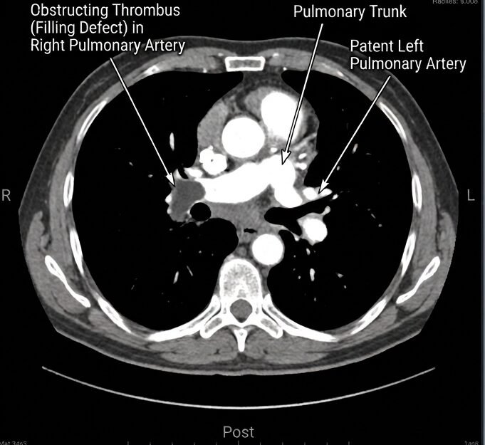
- The undisputed first-line diagnostic gold standard for acute Pulmonary Embolism.
- High Specificity (83%) and High Sensitivity (96%).
- Directly visualizes intraluminal filling defects in main, lobar, segmental, and subsegmental branches.
- Enables simultaneous assessment of right ventricular strain by calculating the RV/LV diameter ratio on axial or four-chamber reformatted views (an RV / LV ratio ≥ 1.0 indicates significant right ventricular enlargement).
- Another advantage of this method is its ability to image and diagnose conditions that mimic Pulmonary Embolism from a clinical perspective.
- Contraindications: patients with renal failure and a history of allergy to the radiographic contrast agent should not undergo CTPA imaging.
Ventilation-Perfusion (V/Q) Scintigraphy:
- Indicated when CTPA is contraindicated (severe renal impairment with eGFR < 30 mL/min/1.73m²), severe iodinated contrast allergy, or pregnant patients with normal baseline chest radiographs.
- It is less effective in diagnosing Pulmonary Embolism compared to CTPA, reaching a diagnostic percentage of 30%-50% of the patients.
- Diagnostic hallmark: Mismatched defects (areas of normal ventilation with absent perfusion, reflecting vascular occlusion). Reported as high, intermediate, or low probability using the modified PIOPED criteria.
Transthoracic Echocardiography (TTE):
- First-line diagnostic tool in hemodynamically unstable, critically ill patients who cannot be safely moved to a CT scanner, making it very valuable in the hospital environment, especially in the emergency department.
- Classic findings: Right ventricular hypokinesis and dilation (RV/LV > 1.0), flattening of the interventricular septum, tricuspid annular plane systolic excursion (TAPSE < 16 mm), and McConnell’s Sign (akinesis of the mid-free Right Ventricle wall with normal or hyperdynamic motion of the RV apex, highly specific for acute Pulmonary Embolism).
Compression Ultrasonography (CUS) of the Lower Extremities:
- Non-invasive duplex imaging of the proximal lower limb deep veins.
- Demonstration of a non-compressible venous segment confirms Deep Vein Thrombosis (normal, non-obstructed veins can typically be pressed with the sound projector of the ultrasonographer so that the circulation inside them can stop). In a patient with suspected PE and compatible cardiopulmonary symptoms, a positive proximal CUS provides sufficient indirect confirmation to initiate full therapeutic anticoagulation without requiring further thoracic imaging.
- Compression Ultrasonography can be useful in pregnant women with a possible Pulmonary Embolism, as well as patients in whom CTPA is contraindicated.
8. Pulmonary Embolism Treatment and Therapeutic Management
Section Summary:
Management of pulmonary embolism hinges on immediate hemodynamic risk stratification. Hemodynamically stable patients receive targeted anticoagulation using direct oral anticoagulants, low-molecular-weight heparin, or fondaparinux to halt thrombus propagation. In high-risk, hemodynamically unstable patients, emergent systemic thrombolysis represents first-line therapy to restore perfusion. When thrombolytics fail or are strictly contraindicated, interventional catheter-directed thrombectomy or surgical pulmonary embolectomy serves as definitive mechanical life-saving revascularization.

1. Acute Resuscitation and Hemodynamic Optimization
In patients with acute PE presenting with shock, initial resuscitation must prioritize supporting right ventricular perfusion while avoiding worsening right heart overload.
Cautious Fluid Administration: Large crystalloid boluses are contraindicated. Over-distending the right ventricle worsens wall tension, increases tricuspid regurgitation, and further displaces the interventricular septum into the left ventricle. If given at all, fluids should be limited to a single conservative challenge (≤ 500 mL of balanced crystalloid over 20–30 minutes).
Vasopressor and Inotropic Support:
- Norepinephrine (0.05 μg/kg/min) is the first-line vasopressor. It stimulates peripheral α1-adrenergic receptors to raise aortic root diastolic pressure (restoring right coronary artery perfusion), while its modest β1-adrenergic activity improves Right Ventricle myocardial inotropy.
- Dobutamine (2 – 10 μg/kg/min) can be added to improve forward cardiac output in low-flow states, but should be titrated cautiously to avoid tachycardia or peripheral vasodilation.
Oxygenation and Mechanical Ventilation:
- Deliver supplemental oxygen via nasal cannula or high-flow nasal cannula (HFNC) to maintain SpO2 ≥ 90%.
- Endotracheal Intubation and mechanical ventilation should be avoided if possible. The induction agents can cause systemic vasodilation, and the shift to positive-pressure ventilation increases intrathoracic pressure, suddenly decreasing venous return (RV preload) and precipitating circulatory collapse. If intubation is unavoidable, use hemodynamically stable induction agents (e.g., ketamine or etomidate) and maintain low tidal volumes (6 ml/kg) with low positive end-expiratory pressure (PEEP).
2. Pharmacological Anticoagulation
Anticoagulation is the cornerstone of therapy for all patients with acute Pulmonary Embolism (PE). It does not actively dissolve existing clots; rather, it halts thrombus propagation, prevents recurrent embolic showers, and allows the endogenous fibrinolytic system to gradually clear the occlusion.
Parenteral Anticoagulants:
-Unfractionated Heparin (UFH):
- Mechanism: Binds antithrombin III, accelerating inactivation of thrombin (Factor IIa) and Factor Xa.
- Dosing: Intravenous bolus of 80 units/kg, followed by a continuous infusion of 18 units/kg/h, titrated to a target activated partial thromboplastin time (aPTT) of 1.5 to 2.5 times control (or anti-Xa level 0.3–0.7 IU/mL).
- Preferred Indications: Classic unfractionated heparin is used only in patients with hemodynamic instability (high-risk PE patients who cannot undergo thrombolysis), severe renal insufficiency (< 30 ml/min/1.73 m²), extreme obesity, or high anticipated bleeding risk where rapid reversal with protamine sulfate can be done if needed.
-Low-Molecular-Weight Heparin (LMWH):
- Agents: Enoxaparin (1 mg/kg subcutaneously every 12 hours or 1.5 mg/kg once daily) or Dalteparin (200 units/kg once daily).
- Advantages: Predictable pharmacokinetics, higher bioavailability, no requirement for routine laboratory monitoring, and significantly lower incidence of Heparin-Induced Thrombocytopenia (HIT). Requires dose reduction or monitoring of anti-Xa levels in moderate renal impairment.
-Fondaparinux:
- A synthetic pentasaccharide that selectively inhibits Factor Xa. Administered subcutaneously once daily based on weight (5 mg if < 50 kg, 7.5 mg if 50 – 100 kg, 10 mg if > 100 kg). Contraindicated if Creatinine Clearance < 30 ml/min.
Direct Oral Anticoagulants (DOACs)
DOACs represent the first-line oral anticoagulant therapy for hemodynamically stable patients with acute Pulmonary Embolism (intermediate- and low-risk), preferred over Vitamin K antagonists (warfarin) due to lower risks of major and intracranial hemorrhage, rapid onset of action, and no need for routine INR monitoring.
- Rivaroxaban (Oral Factor Xa Inhibitor): Monotherapy, 15 mg orally twice daily with food for the first 21 days, followed by 20 mg orally once daily thereafter.
- Apixaban (Oral Factor Xa Inhibitor): Monotherapy, 10 mg orally twice daily for the first 7 days, followed by 5 mg orally twice daily thereafter.
- Dabigatran (Direct Thrombin / Factor IIa Inhibitor): Requires a parenteral lead-in (LMWH or UFH) for at least 5 days, followed by 150 mg orally twice daily.
- Edoxaban (Oral Factor Xa Inhibitor): Requires a parenteral lead-in (LMWH or UFH) for at least 5 days, followed by 60 mg orally once daily (reduced to 30 mg if creatinine clearance is 15 – 50 mL/min or body weight ≤ 60 kg).
Vitamin K Antagonists (Warfarin)
Reserved for patients with severe renal impairment (creatinine clearance < 15 mL/min), antiphospholipid syndrome (APS, where DOACs are contraindicated due to higher rates of arterial recurrence), mechanical heart valves, or when DOACs are financially inaccessible.
- Must be co-administered with a parenteral anticoagulant (LMWH or UFH) for a minimum of 5 days and until the International Normalized Ratio (INR) is therapeutic (INR 2.0 – 3.0) for at least two consecutive days.
Pharmacological Treatment in Pulmonary Embolism Overview – Duration of therapy
The duration of Pulmonary Embolism therapy does not exceed three months for patients who have a reversible risk factor (estrogen therapy, surgery, trauma, temporary immobilization, pregnancy). On the contrary, long-term therapy for Pulmonary Embolism should be assessed in every patient with idiopathic Pulmonary Embolism who does not present a high risk of bleeding. Finally, in patients with neoplasms or high-risk thrombophilia (such as positive lupus anticoagulant, protein C deficiency, protein S deficiency, homozygous for the factor V Leiden, prothrombin mutation, G20210A), the anticoagulation therapy should be continued indefinitely.
3. Systemic Thrombolytic Therapy
Indicated for patients presenting with High-Risk (Massive) Pulmonary Embolism accompanied by persistent systemic hypotension, cardiogenic shock, or cardiopulmonary arrest. Thrombolysis offers clearer advantages within the first 48 hours after symptom onset but remains effective for 6 to 14 days after. Thrombolysis directly converts plasminogen to active plasmin, accelerating enzymatic degradation of the clot and rapidly unloading the right ventricle.
- Regimen: Recombinant tissue plasminogen activator (Alteplase / rtPA) 100 mg administered as a continuous intravenous infusion over 2 hours. In extreme cardiac arrest scenarios, an accelerated regimen of 0.6 mg/kg (maximum 50 mg) infused over 15 minutes can be used. Recombinant tissue plasminogen (rtPA) is the newer medication. Other medications include streptokinase and urokinase.
Absolute Contraindications to Thrombolysis:
- Prior history of any hemorrhagic stroke or stroke of unknown origin.
- Ischemic stroke within the preceding 6 months.
- Known central nervous system neoplasm or vascular malformation (aneurysm, AVM).
- Recent major trauma, surgery, or head injury within the past 3 weeks.
- Active internal bleeding (excluding menses).
- Known bleeding diathesis.
4. Advanced Interventional and Surgical Modalities
When systemic thrombolysis is strictly contraindicated, has failed, or when the patient is in extremis:
- Catheter-Directed Thrombolysis (CDT):
Delivers low-dose thrombolytic therapy directly into the pulmonary thrombus through endovascular catheters, with or without ultrasound assistance (e.g., the EKOS system). This reduces the total required lytic dose (e.g., 10-20 mg alteplase), lowering the risk of major systemic and intracranial hemorrhage.
- Percutaneous Mechanical Thrombectomy:
Catheter-based aspiration or mechanical maceration devices (e.g., Inari FlowTriever, Penumbra Lightning) that physically extract large-volume thrombi from the central pulmonary arteries without requiring thrombolytic agents.
- Surgical Pulmonary Embolectomy:
Open surgical removal of obstructing emboli via median sternotomy and cardiopulmonary bypass. Indicated in massive Pulmonary Embolism with absolute contraindications to thrombolysis, failed systemic/catheter therapy, or intracardiac thrombi straddling the foramen ovale (“clot-in-transit”).
- Inferior Vena Cava (IVC) Filters:
Mechanical mesh devices deployed endovascularly into the infrarenal IVC.
Strict Indications: Documented acute VTE with an absolute contraindication to anticoagulation (e.g., active intracranial bleeding) or objectively verified recurrent PE despite therapeutic-range anticoagulation. Retrievable filters are preferred, and anticoagulation should be resumed and the filter removed as soon as the bleeding contraindication resolves.
9. Pulmonary Embolism Prognosis, Prevention, Risk Stratification, and Long-Term Sequelae
Section Summary:
Prognosis in pulmonary embolism depends heavily on baseline hemodynamic stability, right ventricular strain, and validated prognostic scores such as the Pulmonary Embolism Severity Index. While prompt anticoagulation limits acute mortality in low-risk disease below two percent, massive embolism carries catastrophic in-hospital mortality. Long-term surveillance remains vital to detect persistent functional disability, post-pulmonary embolism syndrome, and chronic thromboembolic pulmonary hypertension requiring dedicated pulmonary vascular intervention.
Short- and long-term prognosis after acute Pulmonary Embolism (PE) is determined by the severity of acute right ventricular strain and underlying cardiopulmonary reserve.
The Pulmonary Embolism Severity Index (PESI)
The PESI score and its simplified version (sPESI) are internationally validated prognostic tools used to predict 30-day all-cause mortality and identify low-risk candidates for early hospital discharge or outpatient management.
The Simplified PESI (sPESI)
| Clinical Variable | Assigned Points |
| Age > 80 years | 1 point |
| History of Cancer | 1 point |
| Chronic Heart Failure or Chronic Pulmonary Disease | 1 point |
| Pulse rate 110 beats per minute | 1 point |
| Systolic Blood Pressure < 100 mmHg | 1 point |
| Arterial Oxygen Saturation SpO2 < 90% | 1 point |
Risk Interpretation:
- 0 Points: Low 30-day mortality risk (~ 1.0%). Candidate for early discharge and home outpatient treatment with DOAC therapy (provided adequate home social support is available).
- ≥ 1 point: High 30-day mortality risk (~ 10.9). Requires inpatient hospitalization and continuous monitoring.
Acute Mortality Stratification
- High-Risk (Massive) PE: In-hospital mortality exceeds 15% to 30%, with most deaths occurring within the first 1 to 2 hours of clinical presentation due to irreversible right ventricular failure.
- Intermediate-Risk (Submassive) PE: 30-day mortality ranges from 3% to 8%, primarily driven by secondary hemodynamic collapse in intermediate-high-risk patients.
- Low-Risk PE: 30-day mortality is < 1% to 2% when therapeutic anticoagulation is promptly initiated.
Long-Term Sequelae
Post-Pulmonary Embolism Syndrome (PPES): Characterized by chronic, persistent dyspnea, exercise intolerance, and impaired functional quality of life lasting > 3 to 6 months following an acute Pulmonary Embolism, in the absence of recurrent events. This affects up to 30% to 50% of survivors and is driven by deconditioning, residual microvascular perfusion defects, and persistent subtle right ventricular remodeling.
Chronic Thromboembolic Pulmonary Hypertension (CTEPH):
- A serious long-term vascular complication occurring in approximately 1% to 4% of Pulmonary Embolism survivors within 2 years.
- Pathogenesis: Failure of endogenous fibrinolysis leads to fibrotic organization and recanalization of unresolved thrombi within pulmonary arteries. This triggers progressive secondary vascular remodeling in non-occluded vessels, causing severe pulmonary hypertension, progressive right heart failure, and death.
- Diagnosis: Screened with transthoracic echocardiography; confirmed with V/Q scintigraphy (demonstrating mismatched segmental defects) and invasive Right Heart Catheterization showing a mean pulmonary arterial pressure (mPAP > 20 mmHg) with a pulmonary capillary wedge pressure (PCWP ≤ 15 mmHg) and elevated PVR ≥ 2 Wood Units.
- Management: Potentially curable via Pulmonary Thromboendarterectomy (PTE) surgery. For non-surgical candidates, treatment involves balloon pulmonary angioplasty (BPA) and medical therapy with Riociguat (a soluble guanylate cyclase stimulator).
Pulmonary Embolism Prevention
The prevention of pulmonary embolism involves two basic principles:
- Recognizing high-risk patients (look at the sPESI table above).
- Applying effective preventive treatment.
Special emphasis in preventive measures is given to orthopedic surgeries of the hip and the knee, as well as major surgical procedures in patients over the age of 40 years. For protection, low-weight heparins are used in one or two doses within a 24-hour period without the need for laboratory testing.
10. Pulmonary Embolism Interconnection with Cardiovascular, Renal, and Metabolic Diseases
Section Summary:
Pulmonary embolism shares complex pathophysiological links and shared risk factors with major cardiovascular, renal, and metabolic disorders. Endothelial dysfunction, systemic vascular inflammation, and prothrombotic signaling unite pulmonary embolism with atherosclerosis, coronary artery disease, hypertension, and type 2 diabetes mellitus. Furthermore, pulmonary embolism significantly exacerbates congestive heart failure and atrial fibrillation through acute right ventricular strain, while chronic kidney disease impairs antithrombotic clearance and accelerates hypercoagulability.

Pulmonary embolism does not develop in isolation. It shares physiological triggers, clinical presentations, and therapeutic overlap with multiple core cardiovascular and metabolic diseases.
1. Coronary Artery Disease (CAD) and Atherosclerosis
- Diagnostic Overlap: Acute submassive or massive Pulmonary Embolism often mimics Acute Coronary Syndrome (ACS). Both present with retrosternal chest pain, dyspnea, diaphoresis, and elevated cardiac troponins. In Pulmonary Embolism, troponin release reflects acute Right Ventricle subendocardial strain rather than primary coronary plaque rupture.
- The Cardiovascular Continuum: Landmark epidemiologic studies demonstrate that arterial atherosclerosis and venous thromboembolism share common risk factors, including hyperlipidemia, metabolic syndrome, and smoking. Endothelial dysfunction and systemic vascular inflammation provide a shared biological foundation for both atheroma formation and venous thrombosis.
2. Congestive Heart Failure (CHF)
- Bidirectional Risk: Chronic heart failure is an independent predictor of Pulmonary Embolism (Odds Ratio ~ 2-3). Poor forward cardiac output, low venous flow velocities in the lower extremities, and elevated central venous pressure cause peripheral venous stasis. Concurrently, hepatic congestion impairs clearance of activated clotting factors.
- Catastrophic Hemodynamic Impact: In patients with pre-existing left ventricular failure, an acute pulmonary embolism—even a small subsegmental occlusion—can trigger rapid cardiopulmonary collapse because the patient lacks the cardiopulmonary reserve to tolerate any sudden increase in right ventricular afterload. Both conditions cause dyspnea and elevate BNP/NT-proBNP, making echocardiographic evaluation essential to distinguish isolated left heart failure from acute cor pulmonale.
3. Systemic Arterial Hypertension
- Vascular Cross-Talk: Chronic systemic arterial hypertension drives systemic vascular remodeling, microvascular rarefaction, and arterial stiffness. While systemic hypertension affects the high-pressure left-sided circulation, the associated endothelial injury, chronic low-grade inflammation, and accelerated vascular aging contribute to an overall prothrombotic vascular phenotype.
4. Atrial Fibrillation (AFib)
- Arrhythmogenic Trigger: The acute right atrial distension and elevated intracavitary pressures caused by pulmonary embolism frequently trigger acute supraventricular tachyarrhythmias, particularly new-onset atrial fibrillation or atrial flutter.
- Therapeutic Synergy: Direct oral anticoagulants (apixaban, rivaroxaban, edoxaban, dabigatran) represent the first-line therapeutic standard for both stroke prevention in non-valvular Atrial Fibrillation and the treatment of acute Pulmonary Embolism, creating complete therapeutic overlap in patients with both conditions.
5. Type 2 Diabetes Mellitus (T2DM)
- Prothrombotic State: Type 2 Diabetes Mellitus promotes hypercoagulability through multiple mechanisms: chronic hyperglycemia accelerates non-enzymatic glycation of coagulation factors, elevates Plasminogen Activator Inhibitor-1 (PAI-1) (which blunts endogenous fibrinolysis), induces endothelial dysfunction via advanced glycation end-products (AGEs), and drives platelet hyperactivity. Patients with Type 2 Diabetes Mellitus carry an increased baseline risk for both provoked and unprovoked venous thromboembolism.
6. Chronic Kidney Disease (CKD)
- Complex Coagulopathy: Chronic Kidney Disease patients exist in a delicate, paradoxical state characterized by both accelerated platelet dysfunction (uremic bleeding) and systemic hypercoagulability (chronic inflammation, loss of natural anticoagulants in proteinuria).
- Diagnostic and Pharmacological Challenges: In advanced CKD (eGFR < 30 ml/min/1.73 m²), iodinated contrast agents for CTPA carry a risk of contrast-induced nephropathy, necessitating alternative modalities like V/Q scans. Furthermore, low-molecular-weight heparins and DOACs rely on renal elimination to varying degrees, requiring dose adjustments or a switch to unfractionated heparin and warfarin to avoid toxic drug accumulation.
11. Frequently Asked Questions (FAQs)
Section Summary:
Frequently asked questions about pulmonary embolism address critical concerns regarding recurrence risk, recovery timelines, physical limitations during therapy, travel precautions, and the role of blood-thinning medications. Understanding these clinical aspects empowers patients to identify recurrent thrombotic warning signs early, manage ongoing anticoagulant therapies safely, mitigate bleeding complications, and adopt practical lifestyle modifications that substantially improve long-term functional recovery and reduce cardiovascular morbidity.
1. How long do I need to take blood thinners (anticoagulants) after a pulmonary embolism?
The duration of anticoagulation depends on whether the Pulmonary Embolism was provoked by a temporary risk factor or was unprovoked:
- Transient / Provoked Pulmonary Embolism (e.g., following major surgery, bone fracture, or temporary immobility): Standard guidelines recommend 3 months of therapeutic anticoagulation. Once the provoking factor has fully resolved, therapy can safely be discontinued.
- First Unprovoked Pulmonary Embolism: A minimum of 3 to 6 months is mandatory, followed by an evaluation of long-term risks and benefits. If the patient has a low bleeding risk, extended or indefinite anticoagulation is often recommended because stopping treatment carries a recurrence risk of up to 30% over 5 years.
- Recurrent Pulmonary Embolism or Persistent Risk Factors (e.g., active cancer, Antiphospholipid Syndrome): Requires indefinite anticoagulation, with periodic reassessments of bleeding risk and liver/kidney function.
2. Can a pulmonary embolism dissolve completely on its own?
While the human body possesses an innate fibrinolytic system capable of slowly breaking down clots, an acute Pulmonary Embolism of significant size cannot be relied upon to resolve safely without treatment. Without medical intervention, untreated Pulmonary Embolism carries an acute mortality rate approaching 30%. Anticoagulant therapy prevents the clot from propagating or showering new emboli to the lungs, allowing endogenous enzymes to safely dissolve the existing thrombus over weeks to months.
3. What are the earliest warning signs that a pulmonary embolism is recurring?
The earliest signs of recurrence mirror the initial presentation: sudden, unexplained shortness of breath (especially on mild exertion), sharp chest pain that worsens with deep breathing or coughing, an elevated resting heart rate, unexplained lightheadedness, or new coughing with blood-tinged sputum. Patients should also watch for new signs of deep vein thrombosis in the legs, including unilateral swelling, calf pain, tenderness along deep veins, or localized warmth and erythema.
4. Is it safe to exercise or return to work after recovering from a pulmonary embolism?
Return to physical activity should be gradual and supervised. During the first 1 to 2 weeks of anticoagulation, light walking is encouraged to stimulate venous return and reduce stasis. High-intensity aerobic workouts, heavy weightlifting, and contact sports (e.g., martial arts, soccer, basketball) must be avoided while on anticoagulants due to the risk of internal bleeding or traumatic hemorrhage. Return to sedentary work is often feasible within 2 to 4 weeks, while physically demanding jobs may require 2 to 3 months of recovery and formal cardiopulmonary exercise testing.
5. Can I travel by airplane after having a pulmonary embolism?
Air travel is generally discouraged during the first 2 to 4 weeks following an acute PE due to the risks of prolonged sitting, mild hypobaric hypoxia, and dehydration. Once a patient is hemodynamically stable and therapeutic on anticoagulants, travel is safe. For flights lasting longer than 4 hours:
- Stay well hydrated and avoid excessive caffeine or alcohol.
- Perform seated calf-raise exercises and ankle pumps every 30 minutes.
- Walk down the aisle every 1 to 2 hours.
- Wear properly fitted below-knee graduated compression stockings (15–20 or 20–30 mmHg).
12. Essential Questions to Ask Your Doctor
Section Summary:
Asking targeted clinical questions allows patients and families to actively participate in post-pulmonary embolism care. Inquiring about exact anticoagulation duration, necessary laboratory monitoring, bleeding warning signs, genetic thrombophilia testing, and physical rehabilitation parameters ensures clear therapeutic goals. These informed discussions prevent accidental medication errors, guide safe transition back to daily activities, and establish appropriate clinical follow-up for monitoring potential cardiopulmonary complications or long-term chronic vascular strain.

“Was my pulmonary embolism considered ‘provoked’ by a specific trigger, or was it ‘unprovoked’?”
- Clinical Purpose: Identifies the underlying mechanism and helps determine whether anticoagulation should last for 3 months or be continued indefinitely.
“Did the blood clot cause any strain, dilation, or injury to the right side of my heart?”
- Clinical Purpose: Clarifies whether imaging or biomarkers showed right ventricular dysfunction, which informs short-term recovery expectations and post-discharge monitoring.
“What specific direct oral anticoagulant (DOAC) am I being prescribed, and does it require food or kidney function monitoring?”
- Clinical Purpose: Clarifies drug-specific instructions (e.g., rivaroxaban must be taken with a substantial meal; dabigatran requires specific capsule handling; apixaban requires twice-daily dosing) and establishes required renal function check-ups.
“What specific signs of internal or external bleeding should prompt an immediate emergency room visit?”
- Clinical Purpose: Distinguishes minor bleeding (e.g., minor gingival bleeding, small bruises) from major complications requiring immediate intervention (e.g., black tarry stools, coffee-ground emesis, gross hematuria, persistent epistaxis, or sudden severe headache).
“Should I undergo genetic or laboratory testing for an inherited blood clotting disorder (thrombophilia)?”
- Clinical Purpose: Clarifies whether specialized testing is indicated. (Thrombophilia testing is rarely recommended during acute thrombosis or while on anticoagulants, but may be considered weeks after completing therapy in young patients with unprovoked events or strong family histories).
“When should I have a follow-up echocardiogram to evaluate my heart pressures and rule out Chronic Thromboembolic Pulmonary Hypertension (CTEPH)?”
- Clinical Purpose: Establishes a formal timeline (typically 3 to 6 months post-discharge) to screen for unresolved clots and persistent pulmonary vascular strain.
13. Clinically Connected Conditions and Internal Resources
Section Summary:
Exploring the interconnected network between pulmonary embolism and comorbid cardiovascular, metabolic, and systemic disorders is vital for comprehensive clinical management. This internal reference hub connects pulmonary embolism to existing UncutMed clinical guides, including coronary artery disease, heart failure, hypertension, atrial fibrillation, type 2 diabetes, chronic kidney disease, atherosclerosis, and diagnostic blood tests, highlighting shared vascular pathophysiology, overlapping therapeutic strategies, and coordinated disease prevention.
To better understand how pulmonary embolism interacts with broader cardiovascular, metabolic, and systemic diseases, explore these related clinical guides from the UncutMed library:
- [Coronary Artery Disease (CAD): Clinical Pathophysiology, Presentation, and Management]
- Explore the critical differential diagnosis between acute coronary ischemia and pulmonary embolism, including how to distinguish acute right ventricular strain from left anterior descending coronary artery occlusion.
- [Congestive Heart Failure (CHF): Systolic and Diastolic Dysfunction]
- Review how chronic left-sided heart failure causes venous stasis and predisposes to thromboembolism, and how acute pulmonary embolism can precipitate rapid cor pulmonale.
- [Systemic Arterial Hypertension: Pathogenesis, Complications, and Treatment]
- Learn about vascular remodeling, endothelial shear stress, and how systemic vascular disease interacts with low-pressure pulmonary circulation dynamics.
- [Atrial Fibrillation (AFib): Mechanisms, Stroke Prevention, and Anticoagulation]
- Understand how right atrial dilation from acute PE triggers atrial tachyarrhythmias, and examine the overlapping clinical use of DOACs across both conditions.
- [Type 2 Diabetes Mellitus: Endothelial Dysfunction and the Prothrombotic State]
- Examine the cellular mechanisms linking chronic hyperglycemia, PAI-1 elevation, and platelet hyperactivity to increased venous and arterial thrombosis risk.
- [Chronic Kidney Disease (CKD): Pathophysiology and Cardiovascular Complications]
- Explore the complex coagulopathies of renal failure, contrast-induced nephropathy considerations during CTPA, and dosing adjustments for anticoagulants.
- [Atherosclerosis: Plaque Biology, Systemic Inflammation, and Vascular Occlusion]
- Investigate the shared inflammatory mechanisms uniting arterial atherothrombosis and venous thromboembolism across the cardiovascular continuum.
- [Blood Test Explanation: Complete Blood Count, Metabolic Panels, and Cardiac Biomarkers]
- Review the diagnostic utility, normal ranges, and clinical interpretation of D-dimers, troponins, natriuretic peptides (BNP), and serum creatinine.
14. High-Trust Peer-Reviewed References, Sources, and Clinical Guidelines
Section Summary:
This comprehensive clinical guide is grounded in current international cardiopulmonary guidelines and high-impact peer-reviewed medical literature. Recommendations reflect authoritative clinical practice consensus from the European Society of Cardiology, the American Heart Association, the American College of Chest Physicians, and prestigious journals including The New England Journal of Medicine and The Lancet, ensuring clinicians and patients receive rigorously verified, evidence-based diagnostic criteria and contemporary therapeutic standards.
- Konstantinides, S. V., Meyer, G., Becattini, C., et al. (2020). 2019 ESC Guidelines for the diagnosis and management of acute pulmonary embolism developed in collaboration with the European Respiratory Society (ERS). European Heart Journal, 41(4), 543–603. DOI: 10.1093/eurheartj/ehz405.
- Stevens, S. M., Woller, S. C., Kreuziger, L. M., et al. (2021). Executive Summary: Antithrombotic Therapy for VTE Disease: CHEST Guideline and Expert Panel Report. CHEST, 160(6), 2247–2259. DOI: 10.1016/j.chest.2021.07.056.
- Jaff, M. R., McMurtry, M. S., Archer, S. L., et al. (2011). Management of Massive and Submassive Pulmonary Embolism, Iliofemoral Deep Vein Thrombosis, and Chronic Thromboembolic Pulmonary Hypertension: A Scientific Statement From the American Heart Association. Circulation, 123(16), 1788–1830. DOI: 10.1161/CIR.0b013e318214914f.
- Wells, P. S., Anderson, D. R., Rodger, M., et al. (2000). Derivation of a simple clinical model to categorize patients’ probability of pulmonary embolism: Increasing the utility of the SIMPLIFY D-dimer assay. Thrombosis and Haemostasis, 83(3), 416–420.
- Kline, J. A., Mitchell, A. M., Kabrhel, C., et al. (2004). Clinical criteria to prevent unnecessary diagnostic testing in emergency department patients with suspected pulmonary embolism (The PERC Rule). Journal of Thrombosis and Haemostasis, 2(8), 1247–1255.
- Agnelli, G., Becattini, C., Bauersachs, R., et al. (2013). Oral rivaroxaban for the treatment of symptomatic pulmonary embolism: The EINSTEIN-PE study. The New England Journal of Medicine, 366(14), 1287–1297.
- Agnelli, G., Buller, H. R., Cohen, A., et al. (2013). Oral apixaban for the treatment of acute venous thromboembolism: The AMPLIFY study. The New England Journal of Medicine, 369(9), 799–808.
- Meyer, G., Vicaut, E., Danays, T., et al. (2014). Fibrinolysis for patients with intermediate-risk pulmonary embolism: The PEITHO randomized trial. The New England Journal of Medicine, 370(15), 1402–1411.
- Rali, P. M., & Criner, G. J. (2018). Submassive Pulmonary Embolism. American Journal of Respiratory and Critical Care Medicine, 198(5), 588–598.
- Kim, N. H., Delcroix, M., Jais, X., et al. (2019). Chronic thromboembolic pulmonary hypertension. European Respiratory Journal, 53(1), 1801915. DOI: 10.1183/13993003.01915-2018.
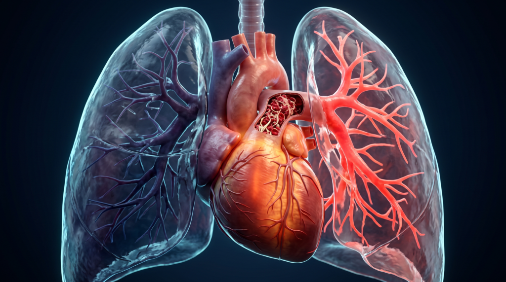
Featured Image: 3D illustration of a thromboembolism at the bifurcation of the pulmonary artery. The image was created with the help of artificial intelligence.
© UncutMed — Medical knowledge, precise, truthful, unfiltered. The current article was last modified on October 18, 2026. This article was clinically authored and reviewed under international evidence-based standards. Always consult your personal healthcare physician before modifying any medical therapy.