© UncutMed — Medical knowledge, precise, truthful, unfiltered. The current article was last modified on October 24, 2026. This article was clinically authored and reviewed under international evidence-based standards. Always consult your personal healthcare physician before modifying any medical therapy.
Medical Disclaimer: This article is for informational and educational purposes only. It does not constitute medical advice, formal diagnosis, or treatment recommendations. Always consult a qualified healthcare professional regarding any metabolic condition or therapeutic plan.
1. Iron Deficiency Anemia Clinical Definition
Section Summary:
Iron deficiency anemia (IDA) is a microcytic, hypochromic hematologic disorder characterized by diminished total body iron stores that fail to support physiological hemoglobin synthesis and erythropoiesis. Defined by the World Health Organization as hemoglobin concentrations below age- and sex-specific thresholds paired with depleted serum ferritin, Iron Deficiency Anemia represents the terminal stage of negative iron balance and remains the leading cause of anemia worldwide.
Iron deficiency anemia (IDA) is a quantitative hematologic disorder in which total body iron stores are exhausted to a degree that restricts erythroid progenitor hemoglobinization, resulting in a reduced systemic concentration of circulating erythrocytes and hemoglobin (Hb). Iron is an indispensable transition metal required not only for oxygen transport via the heme prosthetic group of hemoglobin and oxygen storage via myoglobin, but also for mitochondrial oxidative phosphorylation (cytochromes a, b, and c), iron-sulfur (Fe-S) cluster enzymes, DNA replication (ribonucleotide reductase), and neurotransmitter biosynthesis.
Clinically, iron deficiency evolves along a continuum comprising three distinct biochemical stages before manifesting as frank anemia:
- Stage I (Storage Iron Depletion / Negative Iron Balance): Iron losses or physiological demands exceed intestinal absorption. Reticuloendothelial macrophage and hepatocyte ferritin and hemosiderin reserves are mobilized and progressively depleted. Serum ferritin declines below 30 ng/mL, while serum iron, transferrin saturation (TSAT), erythrocyte indices, and hemoglobin concentration remain within normal reference intervals.
- Stage II (Iron-Restricted Erythropoiesis / Non-Anemic Iron Deficiency): Storage pools are exhausted, and circulating transport iron becomes insufficient to saturate bone marrow erythroblast transferrin receptors. Transferrin Saturation drops below 16%, soluble transferrin receptor (sTfR) levels rise, reticulocyte hemoglobin content (CHr or Ret-He) declines below 29 pg, and free erythrocyte protoporphyrin accumulates (incorporating zinc to form zinc protoporphyrin). Total hemoglobin concentration remains above the lower limit of normal, and mean corpuscular volume (MCV) typically stays normocytic.
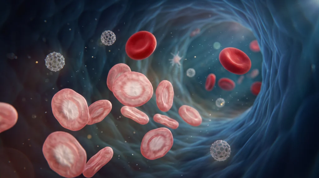
- Stage III (Iron Deficiency Anemia): Persistent iron starvation impairs hemoglobin translation in bone marrow normoblasts, triggering extra mitotic divisions coupled with reduced cytoplasmic volume. The peripheral blood demonstrates frank anemia (defined by the World Health Organization as Hb below 13.0 g/dL in adult males, below 12.0 g/dL in non-pregnant adult females, and below 11.0 g/dL in pregnant individuals) accompanied by classic microcytosis (MCV below 80 fL), hypochromia (mean corpuscular hemoglobin [MCH] below 27 pg), and elevated red cell distribution width (RDW above 14.5%).
2. Iron Deficiency Anemia Causes and Etiology
Section Summary:
The etiology of iron deficiency anemia stems from an imbalance between physiological iron acquisition and systemic iron loss or utilization. In adult males and postmenopausal females, chronic occult gastrointestinal bleeding—frequently secondary to peptic ulceration, angiodysplasia, or colorectal malignancy—is the predominant cause. In premenopausal females, menorrhagia and gestational demands predominate, whereas duodenal malabsorption, dietary insufficiency, and chronic intravascular hemolysis account for remaining cases.
Because humans lack an active physiological excretory pathway for iron—losing only 1 to 2 mg daily via desquamated duodenal enterocytes, cutaneous sloughing, sweat, and biliary/urinary trace losses—the development of Iron Deficiency Anemia in an adult invariably signifies pathological blood loss, defective intestinal mucosal transport, or uncompensated physiological expansion of the erythroid mass.
Chronic Blood Loss (The Predominant Mechanism in Developed Nations)
Each milliliter of packed erythrocytes contains approximately 1 mg of elemental iron (or 0.5 mg per milliliter of whole blood with a normal hematocrit). Consequently, chronic loss of as little as 2 to 4 mL of blood per day exceeds the maximal compensatory capacity of duodenal dietary absorption.
- Gynecological Blood Loss: Heavy menstrual bleeding (menorrhagia, defined clinically as blood loss exceeding 80 mL per cycle) is the single most common cause of Iron Deficiency Anemia in premenopausal women. Structural etiologies include uterine leiomyomas (fibroids), adenomyosis, endometrial polyps, endometrial hyperplasia, and copper intrauterine device (IUD)-induced endometrial inflammation, alongside systemic coagulopathies such as von Willebrand disease.
- Upper Gastrointestinal Tract Bleeding: Esophagitis, gastroesophageal varices, Mallory-Weiss tears, Helicobacter pylori-associated or nonsteroidal anti-inflammatory drug (NSAID)-induced peptic ulcer disease (gastric and duodenal ulcers), Cameron lesions within large hiatal hernias, gastric antral vascular ectasia (GAVE or “watermelon stomach”), portal hypertensive gastropathy, and gastric adenocarcinoma.
- Lower Gastrointestinal Tract Bleeding: Colorectal adenocarcinoma (particularly right-sided cecal and ascending colon neoplasms, which bleed occultly without overt hematochezia), adenomatous polyps, inflammatory bowel disease (ulcerative colitis and Crohn’s disease), colonic angiodysplasia (vascular ectasias, occasionally linked to aortic stenosis in Heyde syndrome), diverticular disease, ischemic colitis, and chronic hemorrhoidal bleeding.
- Small Bowel (Obscure) Bleeding: Meckel diverticulum, jejunoileal angioectasias, gastrointestinal stromal tumors (GISTs), small bowel lymphoma, and NSAID-induced diaphragm disease.
- Helminthic Parasitic Infestations: Globally, hookworm infection (Ancylostoma duodenale and Necator americanus) is a leading cause of Iron Deficiency Anemia. Adult worms attach to the jejunal mucosa, secrete anticoagulants, and ingest host blood; Ancylostoma duodenale causes approximately 0.15 to 0.25 mL of blood loss per worm per day. Trichuris trichiura (whipworm) and Schistosoma mansoni or Schistosoma haematobium (causing intestinal or urinary schistosomiasis, respectively) also drive severe endemic iron depletion.
- Urinary and Pulmonary Blood Loss: Chronic hematuria from renal cell carcinoma, urothelial carcinoma, or IgA nephropathy; pulmonary hemosiderosis and diffuse alveolar hemorrhage (e.g., Goodpasture syndrome, granulomatosis with polyangiitis), where iron is sequestered within alveolar macrophages as hemosiderin and rendered unavailable for erythropoiesis.
- Intravascular Hemolysis and Urinary Iron Loss: Paroxysmal nocturnal hemoglobinuria (PNH), mechanical prosthetic heart valve hemolysis (shear-stress fragmentation), and march hemoglobinuria result in saturation of plasma haptoglobin and hemopexin, leading to persistent hemoglobinuria and hemosiderinuria via shed renal proximal tubular epithelial cells.
- Iatrogenic and Factitious Losses: Frequent diagnostic phlebotomy in critically ill intensive care patients, regular voluntary blood donation (each 450–500 mL unit removes 200–250 mg of elemental iron), chronic hemodialysis circuit blood trapping, and factitious self-phlebotomy (Lasthénie de Ferjol syndrome).
Impaired Gastrointestinal Iron Absorption
Dietary non-heme iron is absorbed almost exclusively in the duodenum and proximal jejunum, requiring an intact mucosal brush border and an acidic gastric environment to reduce insoluble ferric iron (Fe3+) to soluble ferrous iron (Fe2+).
- Celiac Disease (Gluten-Sensitive Enteropathy): Autoimmune villous atrophy of the duodenum impairs enterocyte surface area and downregulates brush-border transport proteins; occult celiac disease accounts for 3% to 5% of unexplained Iron Deficiency Anemia cases presenting without classic steatorrhea.
- Gastric Achlorhydria and Hypochlorhydria: Autoimmune atrophic gastritis (which often precedes pernicious anemia by decades), chronic Helicobacter pylori pangastritis (which consumes ascorbic acid and neutralizes gastric acid via bacterial urease), and long-term pharmacotherapy with proton pump inhibitors (PPIs) or histamine-2 (H2) receptor antagonists prevent the solubilization of dietary ferric salts.
- Bariatric and Foregut Surgery: Roux-en-Y gastric bypass, sleeve gastrectomy, and Billroth II gastrojejunostomy both reduce gastric acid secretion and surgically bypass the primary absorptive enterocytes of the duodenum and proximal jejunum.
- Enteric Mucosal Inflammation: Crohn’s disease involving the duodenum/jejunum, tropical sprue, Whipple disease, and intestinal lymphangiectasia.
Inadequate Dietary Intake
- Nutritional Deprivation: Predominant in socioeconomically disadvantaged regions where staple diets consist heavily of cereal grains and legumes rich in phytates (inositol hexaphosphate) and polyphenols, which chelate non-heme iron in the gut lumen into non-absorbable complexes.
- Restrictive Diets: Poorly supplemented vegan or strict vegetarian diets (lacking highly bioavailable heme iron from animal myoglobin and hemoglobin), extreme fad diets, anorexia nervosa, and chronic alcoholism.
- Infantile Feeding Practices: Exclusive prolonged breastfeeding beyond 6 months without iron-fortified complementary foods, or premature introduction of unmodified cow’s milk before 12 months of age (which has low bioavailable iron, inhibits non-heme iron absorption via high casein and calcium content, and induces occult allergic eosinophilic proctocolitis).
Increased Physiological Demand
- Pregnancy and Lactation: A singleton pregnancy requires approximately 1,000 mg of additional elemental iron to support maternal red blood cell mass expansion (around 450–500 mg), fetal and placental tissue accretion (around 300–350 mg), and peripartum blood loss at delivery (around 200 mg).
- Infancy, Childhood, and Adolescence: Rapid somatic growth spurts and lean muscle mass expansion (increasing myoglobin synthesis) combined with the onset of menarche in adolescent females.
- Erythropoiesis-Stimulating Agent (ESA) Therapy: Administration of recombinant human erythropoietin (epoetin alfa, darbepoetin alfa) in chronic kidney disease or oncology patients accelerates bone marrow erythroid proliferation at a rate that exceeds the kinetics of transferrin-mediated iron mobilization from macrophage stores, inducing functional iron deficiency.
Genetic Causes (Iron-Refractory Iron Deficiency Anemia – IRIDA)
- TMPRSS6 Mutations: An autosomal recessive disorder caused by loss-of-function mutations in the TMPRSS6 gene, which encodes matriptase-2, a hepatocyte transmembrane serine protease. Without functional matriptase-2 to cleave hemojuvelin (HJV), the BMP-SMAD signaling cascade remains constitutively active, driving inappropriately elevated hepatic hepcidin synthesis that blocks intestinal and macrophage ferroportin, resulting in lifelong microcytic anemia refractory to oral iron therapy.
| Etiological Category | Primary Clinical Examples | Core Pathogenetic Mechanism |
| Chronic Blood Loss | Menorrhagia, peptic ulcers, colorectal cancer, hookworm | Direct depletion of circulating hemoglobin-bound iron |
| Malabsorption | Celiac disease, H. pylori gastritis, PPIs, gastric bypass | Loss of duodenal villi, anatomic bypass, or gastric achlorhydria |
| Dietary Insufficiency | Unsupplemented vegan diets, cow’s milk in infants, high-phytate diets | Low intake of bioavailable heme iron or luminal chelation |
| Increased Demand | Pregnancy, lactation, infancy, adolescent growth, ESA therapy | Rapid erythroid and tissue expansion outstripping supply |
| Genetic / Systemic | IRIDA (TMPRSS6 mutation), chronic inflammation (ACD overlap) | Constitutive or cytokine-driven hepcidin elevation blocking ferroportin |
3. Iron Deficiency Anemia Pathophysiology
Section Summary:
Systemic iron homeostasis is governed by the hepatic peptide hormone hepcidin and its cellular receptor, ferroportin. During iron deficiency, suppressed BMP6-SMAD signaling and elevated erythroblast-derived erythroferrone inhibit hepcidin transcription, maximizing duodenal iron absorption via DMT1 and macrophage iron export. When iron depletion persists, reduced intracellular heme activates HRI kinase, phosphorylating eIF2α to halt globin translation and produce microcytic, hypochromic erythrocytes.
The human body contains approximately 3,500 to 4,000 mg of elemental iron in adult males (roughly 50 mg/kg) and 2,000 to 2,500 mg in adult females (roughly 35 to 40 mg/kg). Approximately 65% to 70% of total body iron is incorporated within the hemoglobin of circulating erythrocytes and developing bone marrow normoblasts; 15% to 25% is sequestered within storage complexes (ferritin and hemosiderin) inside reticuloendothelial macrophages of the spleen, liver (Kupffer cells), and bone marrow, as well as hepatic parenchymal cells; 8% to 10% resides in skeletal and cardiac myoglobin; and less than 0.1% (roughly 3 to 4 mg) circulates in plasma bound to the glycoprotein transferrin. Because senescence of erythrocytes after their 120-day lifespan requires the daily synthesis of two hundred billion new red blood cells, the bone marrow demands 20 to 25 mg of elemental iron every 24 hours. Dietary duodenal absorption contributes only 1 to 2 mg/day; the remaining 20 to 24 mg/day is derived from the phagocytic recycling of senescent erythrocytes (erythrophagocytosis) by splenic and hepatic macrophages.
1. Molecular Mechanics of Duodenal Enterocyte Iron Absorption
Dietary iron arrives at the apical brush border of mature duodenal enterocytes in two forms: heme iron (derived from animal hemoglobin and myoglobin, boasting 15% to 35% bioavailability) and non-heme inorganic iron (derived from plants and fortified foods, exhibiting 2% to 10% bioavailability).
- Apical Ferrireduction and Transport: Inorganic dietary iron exists predominantly in the oxidized ferric state (Fe3+), which precipitates at a pH between 3.5 and 3.7. Gastric hydrochloric acid and dietary ascorbic acid (vitamin C) maintain Fe3+ in a soluble, chelated state upon entering the duodenum. At the enterocyte apical microvilli, the membrane-bound ferrireductase duodenal cytochrome b (Dcytb, encoded by CYBRD1) transfers an electron from intracellular ascorbate to reduce (Fe3+) to ferrous iron (Fe2+). The divalent cation is subsequently cotransported across the apical membrane along with a proton (H+) by divalent metal transporter 1 (DMT1, encoded by SLC11A2). Heme iron is internalized intact via apical endocytosis (involving candidate transporters such as heme carrier protein 1 [HCP1] or receptor-mediated endocytic pathways) and degraded within the enterocyte cytosol by heme oxygenase-1 (HO-1) to liberate Fe2+, biliverdin, and carbon monoxide.
- Intracellular Trafficking and Storage: Once inside the enterocyte cytoplasm, Fe2+ enters the labile iron pool (LIP). To prevent Fenton reaction-mediated generation of cytotoxic hydroxyl radicals (H2O2 + Fe2+ → Fe3+ + ·OH + OH−) cytosolic iron chaperones—specifically poly(rC)-binding proteins 1 and 2 (PCBP1 and PCBP2)—bind Fe2+ and deliver it either to ferritin (a 24-subunit spherical protein cage composed of heavy [H] chains with ferroxidase activity and light [L] chains that mineralize up to 4,500 ferric oxyhydroxide ions) or directly to the basolateral exporter. Iron sequestered within enterocyte ferritin is permanently lost into the fecal stream when the enterocyte undergoes apoptosis and exfoliates from the villus tip after 3 to 5 days.
- Basolateral Export and Ferroxidation: Iron destined for systemic circulation is transported across the basolateral membrane of the enterocyte by ferroportin 1 (FPN1, encoded by SLC40A1), the sole known mammalian cellular iron exporter. Because plasma apotransferrin exclusively binds ferric iron (Fe3+), exported (Fe2+) must be immediately oxidized at the basolateral surface by hephaestin (HEPH), a transmembrane multicopper ferroxidase structurally homologous to plasma ceruloplasmin (which performs the equivalent ferroxidase function on macrophages and hepatocytes). Two Fe3+ ions then bind reversibly with high affinity to a single molecule of plasma apotransferrin, forming holotransferrin (diferric transferrin) for delivery to the bone marrow.
2. The Hepcidin-Ferroportin Regulatory Axis
Systemic iron flux is orchestrated by hepcidin, a 25-amino-acid disulfide-rich peptide hormone synthesized and secreted by hepatocytes (encoded by the HAMP gene on chromosome 19). Hepcidin acts as the master negative regulator of systemic iron availability by binding directly to the extracellular loop of ferroportin on duodenal enterocytes, reticuloendothelial macrophages, and hepatocytes. Hepcidin binding occludes the central transport cavity of ferroportin and induces its ubiquitination (via the E3 ubiquitin ligase RNF217), clathrin-mediated endocytosis, and lysosomal proteolysis.
- Transcriptional Upregulation of Hepcidin (Iron Overload & Inflammation): When systemic iron stores and plasma diferric transferrin are abundant, hepatic sinusoidal endothelial cells secrete bone morphogenetic protein 6 (BMP6) and BMP2. These ligands bind to a hepatocyte surface receptor complex consisting of BMP type I (ALK2, ALK3) and type II (BMPR2, ActRIIA) serine/threonine kinase receptors along with the GPI-anchored coreceptor hemojuvelin (HJV). Simultaneously, high plasma diferric transferrin displaces the hemochromatosis protein (HFE) from transferrin receptor 1 (TfR1), allowing HFE to complex with transferrin receptor 2 (TfR2) and HJV. This supercomplex phosphorylates intracellular SMAD1/5/8, which complexes with SMAD4 and translocates to the nucleus to activate HAMP transcription. Independently, during infection, malignancy, or autoimmune states, proinflammatory cytokines—principally interleukin-6 (IL-6)—bind to hepatocyte IL-6 receptors, activating the JAK2-STAT3 signaling pathway to drive HAMP transcription, resulting in the iron sequestration characteristic of anemia of chronic disease (anemia of inflammation).
Transcriptional Suppression of Hepcidin in Iron Deficiency: In iron deficiency, three synergistic molecular mechanisms abolish HAMP transcription to maximize intestinal absorption and macrophage iron release:
- BMP-SMAD Attenuation and Matriptase-2 Activation: Low hepatic iron reduces endothelial BMP6 secretion, while low plasma diferric transferrin destabilizes TfR2. Concurrently, iron deficiency stabilizes the hepatocyte transmembrane serine protease matriptase-2 (TMPRSS6), which proteolytically cleaves membrane-bound hemojuvelin (HJV), silencing the BMP-SMAD1/5/8 pathway.
- The Erythroferrone (ERFE) Pathway: Cellular hypoxia in the renal peritubular interstitial fibroblasts prevents the oxygen- and iron-dependent prolyl hydroxylase domain (PHD) enzymes from hydroxylating hypoxia-inducible factor 2-alpha (HIF-2α). Stabilized HIF-2α dimerizes with HIF-1β and induces exponential transcription of erythropoietin (EPO). EPO binds to EPO receptors (EPOR) on bone marrow erythroblasts, activating the JAK2-STAT5 pathway, which induces erythroblasts to secrete the hormone erythroferrone (ERFE, encoded by FAM132B). ERFE circulates to the liver and directly sequesters BMP6 and BMP2 in the space of Dissé, preventing BMP receptor activation and potently suppressing hepcidin synthesis.
- Local Duodenal HIF-2α Stabilization: Within the iron-starved duodenal enterocyte itself, reduced intracellular iron inhibits prolyl hydroxylase 2 (PHD2), stabilizing mucosal HIF-2α, which binds directly to hypoxia-responsive elements (HREs) in the promoters of CYBRD1 (Dcytb), SLC11A2 (DMT1), and SCL40A1 (ferroportin), dramatically upregulating the entire absorptive machinery.
3. Post-Transcriptional Cellular Regulation: The IRE/IRP System
At the individual cellular level, post-transcriptional expression of iron metabolism proteins is regulated by cytosolic iron regulatory proteins 1 and 2 (IRP1 and IRP2), which bind to stem-loop structures termed iron-responsive elements (IREs) located in the untranslated regions (UTRs) of target mRNAs.
- In iron-replete conditions, IRP1 binds a cubane [4Fe-4S] cluster and functions as cytosolic aconitase (unable to bind RNA), while IRP2 undergoes iron-dependent ubiquitination by the FBXL5 E3 ligase complex and proteasomal degradation.
In iron deficiency, the [4Fe-4S] cluster disassembles, and FBXL5 is destabilized, allowing both IRP1 and IRP2 to bind with high affinity to IREs:
- Binding to 5′ UTR IREs (Translational Repression): IRPs binding to the 5′ UTR sterically block recruitment of the 43S pre-initiation ribosomal complex, halting the translation of H- and L-ferritin (preventing iron storage), ferroportin (in erythroid precursors), erythroid 5-aminolevulinate synthase (ALAS2) (preventing toxic protoporphyrin accumulation when iron is absent), and HIF-2α.
- Binding to 3′ UTR IREs (mRNA Stabilization): IRPs binding to the multiple IREs in the 3′ UTR of transferrin receptor 1 (TfR1) and DMT1 mRNAs shield the transcripts from endonucleolytic cleavage by RNase-1 (ZC3H12A), extending mRNA half-life and dramatically increasing cell-surface TfR1 and DMT1 expression to scavenge every available iron atom.
4. Erythroblast Hemoglobinization, HRI Kinase, and the Genesis of Microcytosis
Developing bone marrow erythroblasts (often arranged in erythroblastic islands surrounding a central nurse macrophage) acquire iron exclusively via TfR1-mediated endocytosis of diferric transferrin. Following clathrin-coated pit internalization, vacuolar (H+)-ATPase acidifies the endosome to pH 5.5, causing conformational release of Fe3+ from transferrin. The endosomal ferrireductase STEAP3 (six-transmembrane epithelial antigen of the prostate 3) reduces Fe3+ to Fe2+, which exits the endosome via DMT1 and is imported across the inner mitochondrial membrane via mitoferrin-1 (SLC25A37).
Inside the mitochondrial matrix, the terminal step of heme biosynthesis is catalyzed by ferrochelatase (FECH), a [2Fe-2S] cluster-containing enzyme that inserts Fe2+ into the tetrapyrrole ring of protoporphyrin IX to yield heme. When mitochondrial Fe2+ is depleted in iron deficiency, ferrochelatase substitutes divalent zinc (Zn2+) for iron, generating zinc protoporphyrin (ZPP)—a fluorescent biochemical hallmark of iron-deficient erythropoiesis.
To prevent the proteotoxic aggregation of unpaired α- and β-globin chains when heme is scarce, erythroid precursors rely on heme-regulated inhibitor (HRI) kinase (encoded by EIF2AK1):
- Under normal heme-replete conditions, heme binds directly to the kinase domain of heme-regulated inhibitor (HRI), maintaining it in an inactive conformation.
- In iron deficiency, declining intracellular heme dissociates from HRI, triggering HRI autophosphorylation and activation. Activated HRI phosphorylates the alpha subunit of eukaryotic initiation factor 2 (eIF2α) on serine-51.
- Phosphorylated eIF2α competitively inhibits the guanine nucleotide exchange factor eIF2B, globally attenuating ribosomal translation of α- and β-globin mRNAs so that globin synthesis remains strictly stoichiometric with available heme.
- Simultaneously, the phosphorylated eIF2α / ATF4 integrated stress response alters cell-cycle regulators (such as cyclin D3 and E2F-2) in polychromatophilic normoblasts. Because erythroblasts continue to undergo mitotic cell divisions until a critical intracellular hemoglobin concentration threshold (approximately 20% to 34% MCHC) is sensed, delayed hemoglobin accumulation causes the normoblasts to undergo one or two extra mitotic divisions alongside reduced cytoplasmic growth, yielding mature erythrocytes that are structurally small (microcytic) and hemoglobin-poor (hypochromic).
- Furthermore, iron deficiency increases erythrocyte membrane oxidative stress (due to reduced catalase and glutathione peroxidase activity) and diminishes membrane deformability, inducing poikilocytosis (pencil cells or elliptocytes) and accelerating splenic macrophage clearance, modestly shortening erythrocyte lifespan.
4. Iron Deficiency Anemia Symptoms
Section Summary:
The clinical symptoms of iron deficiency anemia arise from two distinct pathophysiological mechanisms: diminished systemic oxygen delivery to metabolically active tissues due to reduced hemoglobin concentration, and non-hematologic depletion of iron-dependent mitochondrial enzymes and monoamine neurotransmitters. Patients classically present with progressive exertional dyspnea, profound fatigue, cognitive brain fog, restless legs syndrome, and pagophagia—a compulsive craving for ice.
Crucially, many tissue-level symptoms manifest during non-anemic iron deficiency (Stages I and II) before hemoglobin falls below diagnostic thresholds, reflecting the depletion of iron-sulfur clusters and heme enzymes in skeletal muscle and central nervous system neurons. The symptoms of anemia are neither sensitive nor specific, and they do not help distinguish between the many different types of anemia. Symptoms reflect compensatory responses that the body initiates to combat tissue hypoxia and usually develop when the hemoglobin levels fall greatly below the patient’s individual baseline. Symptoms are generally more pronounced in patients with limited cardiopulmonary reserve (such as patients with heart or pulmonary medical conditions) or when the anemia develops extremely quickly.
Systemic and Cardiopulmonary Hypoxic Symptoms
- Chronic Fatigue and Generalized Weakness: The earliest and most ubiquitous complaint, resulting from both impaired oxygen transport and depletion of skeletal muscle myoglobin and mitochondrial cytochrome c oxidase (Complex IV), which shifts muscle metabolism toward early anaerobic glycolysis and lactic acid accumulation.
- Exertional Dyspnea and Reduced Exercise Tolerance: Shortness of breath initially triggered by moderate physical exertion, climbing stairs, or brisk walking, progressing to dyspnea at rest in severe anemia (Hb below 7.0 g/dL).
- Palpitations and Awareness of Heartbeat: Subjective perception of a pounding or racing heart, driven by compensatory sympathetic nervous system activation and an increased hyperdynamic stroke volume and heart rate required to maintain tissue oxygen delivery (DO2).
- Orthostatic Lightheadedness, Presyncope, and Vertigo: Postural dizziness upon standing due to blunted peripheral vasoconstrictive reserve and cerebral hypoperfusion.
- Pulsatile Tinnitus: A rhythmic “whooshing” sound in the ears synchronous with the arterial pulse, caused by turbulent, low-viscosity hyperdynamic blood flow through the petrous carotid canal and jugular bulb.
- Ischemic Chest Pain (Angina Pectoris) or Intermittent Claudication: In patients with pre-existing coronary artery disease or peripheral arterial disease, reduced arterial oxygen content (CaO2) precipitates myocardial or lower-extremity ischemia at lower workloads.
Neuropsychiatric and Neuromuscular Symptoms
- Pica and Pagophagia: Pica is the compulsive ingestion of non-nutritive substances such as clay or soil (geophagia), laundry or cornstarch (amylophagia), chalk, paper, or uncooked pasta. Pagophagia—the compulsive chewing and consumption of large quantities of ice—is highly specific for iron deficiency (present in up to 50% of iron-deficient patients) and usually resolves within days of initiating iron supplementation, long before hemoglobin rises. Mechanistically, chewing ice is hypothesized to trigger a facial dive reflex or local vasoconstriction that increases perfusion to the anterior cingulate cortex of the brain, compensating for sluggish iron-dependent dopamine neurotransmission, helping clear the mental fog and fatigue caused by low iron. Pagophagia also helps relieve the symptoms in the mouth area (such as glossodynia; please read below for further information).
- Restless Legs Syndrome (Willis-Ekbom Disease): An irresistible urge to move the lower extremities accompanied by uncomfortable paresthesias (“creeping” or “crawling” sensations) that worsen during evening rest and are relieved by movement. It stems from regional iron depletion in the substantia nigra and basal ganglia, which impairs tyrosine hydroxylase (an iron-dependent rate-limiting enzyme in dopamine synthesis) and alters striatal D2 dopamine receptor density.
- Cognitive Impairment and “Brain Fog”: Deficits in sustained attention, working memory, executive processing speed, and academic or occupational performance, linked to impaired oligodendrocyte myelination and reduced monoamine (dopamine, norepinephrine, and serotonin) synthesis.
- Chronic Headaches and Irritability: Generalized tension-type or throbbing vascular headaches secondary to cerebral vasodilation in response to reduced arterial oxygen content.
- Sleep Disturbances and Periodic Limb Movement Disorder: Fragmented nocturnal sleep architecture and daytime somnolence.
- Cold Intolerance: Impaired non-shivering thermogenesis and cutaneous vasoconstriction due to defective iron-dependent thyroid peroxidase (TPO) activity, which blunts peripheral conversion of thyroxine (T4) to triiodothyronine (T3).
Epithelial and Oropharyngeal Symptoms
- Glossodynia (Sore, Burning Tongue): Pain, tenderness, or a burning sensation of the tongue, particularly upon consuming acidic or spicy foods, caused by rapid-turnover epithelial atrophy of the lingual filiform papillae.
- Odynophagia and Solid-Food Dysphagia: Progressive difficulty swallowing solids due to post-cricoid esophageal mucosal atrophy and web formation (Plummer-Vinson or Paterson-Brown-Kelly syndrome).
- Hair Loss (Telogen Effluvium) and Brittle Nails: Diffuse scalp hair shedding and painful longitudinal splitting or breaking of the fingernails, reflecting iron inadequacy in the highly proliferative hair follicle matrix (where ribonucleotide reductase requires iron for DNA synthesis).
| Symptom Category | Key Clinical Complaints | Primary Underlying Mechanism |
| Cardiopulmonary | Fatigue, exertional dyspnea, palpitations, angina | Reduced arterial O2 content and mitochondrial cytochrome depletion |
| Neurological | Pagophagia (ice craving), pica, restless legs, brain fog | CNS tyrosine hydroxylase impairment and striatal dopamine dysfunction |
| Epithelial / Mucosal | Sore burning tongue, dysphagia, diffuse hair loss | Impaired ribonucleotide reductase DNA synthesis in rapid-turnover cells |
| Thermoregulatory | Cold intolerance, pale/chilly extremities | Defective thyroid peroxidase (T4 —> T_3$) and peripheral shunting |
5. Iron Deficiency Anemia Signs
Section Summary:
Physical examination in iron deficiency anemia reveals objective clinical signs reflecting both mucocutaneous hypoperfusion and chronic tissue iron starvation. Classic findings include conjunctival and palmar crease pallor, hyperdynamic tachycardia with a systolic flow murmur, koilonychia (spoon-shaped nails), atrophic glossitis with a smooth, depapillated tongue, angular cheilitis, and, in severe chronic cases, post-cricoid esophageal webs characteristic of Plummer-Vinson syndrome.
The physical signs of Iron Deficiency Anemia correlate with both the severity of the hemoglobin deficit and the chronicity of tissue iron depletion.
Mucocutaneous Signs of Anemia
Cutaneous and Mucosal Pallor: Caused by both reduced oxyhemoglobin concentration in the superficial dermal capillary plexus and sympathetic-mediated cutaneous vasoconstriction that shunts blood toward vital organs (heart, brain, kidneys). Because skin pigmentation varies widely, clinical evaluation focuses on non-melanin-bearing vascular beds:
- Palpebral Conjunctival Pallor: Loss of the normal bright red fleshy hue of the anterior rim of the lower tarsal conjunctiva (highly specific when present at Hb levels below 9.0 g/dL).
- Palmar Crease Pallor: Blanching of the deep skin creases of the hyperextended palm (typically evident when Hb falls below 7.0 to 8.0 g/dL).
- Nailbed and Oral Mucosa Pallor: Blanching of the subungual vascular bed and buccal/gingival mucosa.
- Blue Sclerae: A subtle pearly or bluish tint of the ocular sclerae (observed in up to 80% of patients with Iron Deficiency Anemia vs. less than 7% in other anemias). Iron is an essential cofactor for prolyl-4-hydroxylase and lysyl hydroxylase during collagen synthesis; iron deficiency results in thinned, translucent scleral collagen fibers that allow the dark underlying uveal choroid pigment to show through.
Epithelial, Ungual, and Oropharyngeal Signs (Specific to Iron Depletion)
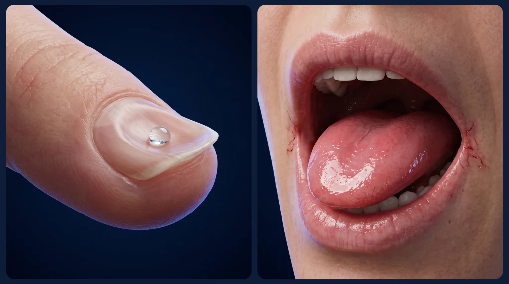
- Koilonychia (Spoon-Shaped Nails): Pathognomonic for chronic severe iron deficiency. The nail plates initially become thin, brittle, and flattened (platonychia), eventually developing a concave central depression with everted lateral margins capable of holding a drop of water on the dorsal surface. This results from impaired cysteine-rich keratin disulfide bond formation and microvascular ischemia of the distal nail matrix.
- Atrophic Glossitis: Patchy or diffuse atrophy and denudation of the filiform and fungiform papillae on the dorsal surface of the tongue, producing a smooth, waxy, glazed, erythematous (“beefy red”) lingual surface.
- Angular Cheilitis (Angular Stomatitis / Perleche): Painful erythematous fissuring, maceration, and ulceration at the labial commissures (corners of the mouth), frequently complicated by secondary colonization with Candida albicans or Staphylococcus aureus.
- Plummer-Vinson (Paterson-Brown-Kelly) Syndrome: The classical clinical triad of chronic iron deficiency anemia, glossitis, and cervical dysphagia caused by thin, eccentric mucosal webs composed of squamous epithelium and lamina propria in the anterior post-cricoid hypopharynx/upper esophagus. It constitutes a premalignant condition associated with a 3% to 15% lifetime risk of post-cricoid or upper esophageal squamous cell carcinoma.
Cardiovascular and Systemic Signs
- Sinus Tachycardia and Bounding Peripheral Pulses (Water-Hammer Pulse Quality): Compensatory elevation in resting heart rate and widened pulse pressure driven by decreased blood viscosity and nitric oxide-mediated peripheral vasodilation.
- Hemic Systolic Flow Murmur: A grade II/VI or III/VI early-to-mid systolic crescendo-decrescendo ejection murmur heard best at the left sternal border or pulmonic valve area, caused by high-velocity, low-viscosity turbulent blood flow across normal semilunar valves.
- Signs of High-Output Heart Failure (in Severe Profound Anemia, Hb below 5.0 g/dL): Cardiomegaly, an audible third heart sound (S3 gallop), elevated jugular venous pressure, bibasilar pulmonary rales, and dependent bilateral pitting ankle edema.
- Anemic Retinopathy: On fundoscopic examination in patients with severe anemia (particularly when Hb is below 6.0 g/dL or accompanied by thrombocytopenia), findings include retinal venous tortuosity, flame-shaped retinal hemorrhages, cotton-wool spots (nerve fiber layer infarcts), and Roth spots (white-centered hemorrhages).
- Splenomegaly: Mild splenomegaly is occasionally palpable in roughly 5% to 10% of chronic, untreated severe Iron Deficiency Anemia cases due to extramedullary erythrophagocytosis of rigid poikilocytes, though prominent splenomegaly should prompt evaluation for coexisting thalassemia, portal hypertension, or lymphoproliferative disease.
6. Iron Deficiency Anemia Risk Factors
Section Summary:
Susceptibility to iron deficiency anemia clusters across distinct physiological, demographic, and pharmacological profiles. Primary risk factors include female reproductive age with heavy menstrual bleeding, multiparity and short interpregnancy intervals, rapid pediatric growth phases, endurance athletics, frequent blood donation, chronic use of nonsteroidal anti-inflammatory drugs or anticoagulants, proton pump inhibitor therapy, and chronic inflammatory or malabsorptive gastrointestinal and renal disorders.
Identifying high-risk populations allows clinicians to implement targeted screening protocols before stage I storage depletion progresses to symptomatic anemia.
Demographic and Physiological Risk Factors
- Menstruating Adolescent and Adult Females: Monthly menstrual blood loss increases basal daily iron requirements from 1 mg/day to approximately 2.0–2.5 mg/day; women using copper IUDs or experiencing menorrhagia face the highest risk.
- Pregnant and Postpartum Individuals: Risk escalates sharply during the second and third trimesters as fetal iron transport across the placenta intensifies; multiparity, twin or multiple gestations, and short interpregnancy intervals (less than 18 months) prevent maternal ferritin recovery between pregnancies.
- Preterm and Low-Birth-Weight Infants: Approximately 80% of fetal iron accretion occurs during the third trimester of gestation via placental transferrin receptors. Infants born prematurely (before 37 weeks) have severely truncated baseline iron stores and experience rapid postnatal catch-up growth.
- Toddlers (Ages 6 to 24 Months) and Adolescents: Periods of peak growth velocity coupled with dietary transitions (such as excessive cow’s milk consumption exceeding 24 ounces/day in toddlers, or fast-food/restrictive diets in teenagers).
- Endurance Athletes (“Runner’s Anemia” / Foot-Strike Hemolysis): Long-distance runners and triathletes experience multifocal iron losses via mechanical intravascular hemolysis in the plantar capillaries during repetitive foot strikes, exercise-induced splanchnic ischemia with occult gastrointestinal microbleeding, hematuria, profuse diaphoresis, and exercise-induced transient spikes in IL-6 and hepcidin 3 to 6 hours post-workout that block post-exercise dietary iron absorption.
- Frequent Voluntary Blood Donors: Donating whole blood three or more times per year in men or two or more times per year in women frequently outpaces dietary iron repletion, leaving up to 35% of regular donors with serum ferritin below 15 ng/mL.
Pharmacological and Dietary Risk Factors
- Chronic NSAID and Aspirin Therapy: Non-selective cyclooxygenase (COX-1 and COX-2) inhibition depletes gastroduodenal mucosal prostaglandin E2 (PGE2), causing mucosal micro-erosions, peptic ulceration, and small-bowel enteropathy.
- Oral Anticoagulant and Dual Antiplatelet Therapy (DAPT): Direct oral anticoagulants (apixaban, rivaroxaban, dabigatran), warfarin, and P2Y12 inhibitors (clopidogrel, ticagrelor) unmask occult gastrointestinal vascular ectasias or colonic neoplasms by provoking subclinical bleeding.
- Chronic Acid-Suppressive Therapy: Long-term use (exceeding 1 to 2 years) of proton pump inhibitors (omeprazole, pantoprazole, esomeprazole) or H2 receptor blockers impairs ferric-to-ferrous iron reduction in the stomach and duodenum.
- High Consumption of Dietary Iron Chelators: Habitual intake of black tea, green tea, coffee (rich in tannins and chlorogenic acid polyphenols), phytate-rich unfermented grains, or high-dose calcium supplements concurrently with meals inhibits non-heme iron absorption by 50% to 70%.
Comorbid Medical and Surgical Risk Factors
- History of Bariatric or Gastric Resection Surgery: Patients post-Roux-en-Y gastric bypass, biliopancreatic diversion, sleeve gastrectomy, or gastrectomy for ulcer/malignancy.
- Chronic Kidney Disease (CKD): Uremic platelet dysfunction causing GI blood loss, chronic hemodialysis blood trapping, and elevated systemic hepcidin (due to both reduced renal clearance of hepcidin and chronic inflammation).
- Congestive Heart Failure (CHF) and Chronic Inflammatory Bowel Disease (IBD): Splanchnic venous congestion and bowel wall edema impair duodenal enterocyte transport, while systemic inflammation upregulates hepcidin.
7. Iron Deficiency Anemia Diagnosis and Diagnostic Criteria
Section Summary:
Diagnosing iron deficiency anemia requires integrating a complete blood count, peripheral blood smear, and comprehensive iron panel. Serum ferritin below 15 ng/mL is virtually 99% specific for absolute iron depletion, whereas thresholds below 30 ng/mL (or below 100 ng/mL with transferrin saturation under 20% in chronic inflammatory states) maximize clinical sensitivity. In men and postmenopausal women, bidirectional endoscopy is mandatory to exclude gastrointestinal malignancy.
A definitive diagnosis of Iron Deficiency Anemia follows a two-step paradigm: (1) biochemical confirmation of iron-deficient erythropoiesis and (2) anatomical identification of the underlying etiology—since Iron Deficiency Anemia is a clinical sign of an underlying pathology rather than a complete diagnosis in itself.
1. Complete Blood Count (CBC) and Red Cell Indices
- Hemoglobin (Hb) and Hematocrit (Hct): Per World Health Organization (WHO) criteria, anemia is defined at sea level as:
-Adult Males (15 years and older): Hb below 13.0 g/dL (130 g/L)
-Non-Pregnant Adult Females (15 years and older): Hb below 12.0 g/dL (120 g/L)
-Pregnant Individuals: Hb below 11.0 g/dL (110 g/L) in the first and third trimesters, and below 10.5 g/dL (105 g/L) in the second trimester (accounting for physiological plasma volume expansion).
- Red Blood Cell (RBC) Count: Decreased proportionally to the hemoglobin concentration (in contrast to β-thalassemia trait, where the Red Blood Cell count is paradoxically normal or elevated due to chronic erythropoietin stimulation).
- Mean Corpuscular Volume (MCV): Classically reduced below 80 fL (microcytic). However, up to 30% to 40% of early Iron Deficiency Anemia cases present with a normocytic MCV (80–100 fL) before complete turnover of the pre-existing normocytic erythrocyte cohort, or when coexisting with folate/vitamin B12 deficiency, creating a dimorphic anemia.
- Mean Corpuscular Hemoglobin (MCH) and Mean Corpuscular Hemoglobin Concentration (MCHC): MCH falls below 27 pg (often the earliest automated index to decline before MCV drops), and MCHC drops below 32 g/dL (hypochromic).
- Red Cell Distribution Width (RDW): Elevated above 14.5%, reflecting marked anisocytosis as newly minted microcytic erythrocytes mix with older normocytic erythrocytes in the peripheral circulation.
- Mentzer Index (MCV/RBC count in millions): A rapid screening ratio to differentiate Iron Deficiency Anemia from thalassemia trait. A Mentzer Index greater than 13 strongly favors Iron Deficiency Anemia (where both MCV and RBC count fall), whereas a value less than 13 favors Thalassemia Trait (where severe microcytosis occurs despite an RBC count that signals erythrocytosis).
- Absolute Reticulocyte Count and Reticulocyte Production Index (RPI): Inappropriately normal or reduced (RPI below 2.0), confirming a hypoproliferative bone marrow defect.
- Platelet Count (Reactive Thrombocytosis): Approximately 15% to 30% of patients with Iron Deficiency Anemia exhibit mild-to-moderate reactive thrombocytosis (platelets exceeding 450.00/μL), driven either by sequence homology between erythropoietin and thrombopoietin or by iron-deficiency-induced biasing of megakaryocytic-erythroid progenitors (MEPs) toward megakaryopoiesis.
2. Peripheral Blood Smear Morphology
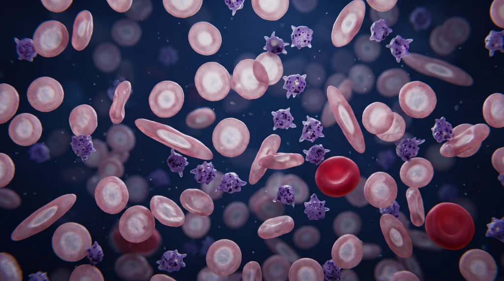
Microscopic inspection of a Wright-Giemsa-stained peripheral blood film in established Iron Deficiency Anemia demonstrates:
- Microcytosis and Hypochromia: Small erythrocytes in which the central zone of pallor exceeds one-third of the total cell diameter (in severe cases appearing as thin peripheral rings of hemoglobin termed anulocytes or “pessary cells”).
- Anisocytosis and Poikilocytosis: Marked variation in red cell size and shape, characteristically featuring elongated cigar- or pencil-shaped cells (elliptocytes) and occasional target cells (codocytes).
- Absence of coarse basophilic stippling (which, when prominent, points toward lead poisoning or thalassemia).
3. Iron Studies and Biochemical Markers
- Serum Ferritin (The Single Most Accurate Non-Invasive Test): Directly correlates with reticuloendothelial storage iron (1 ng/mL} of serum ferritin corresponds to approximately 8 to 10 mg of storage iron).
-Serum Ferritin below 15 ng/mL (μg/L): Per WHO and British Society for Haematology (BSH) guidelines, this confirms absolute iron deficiency with a specificity of 99%.
-Serum Ferritin below 30 ng/mL (μg/L}: Recommended by the American Gastroenterological Association (AGA) and clinical hematology consensus as the optimal diagnostic cutoff in outpatients without inflammation, raising diagnostic sensitivity to 92% while preserving 98% specificity.
-Serum Ferritin in Inflammation, Chronic Kidney Disease, or Heart Failure: Because ferritin is an acute-phase reactant transcriptionally upregulated by IL-1, IL-6, and TNF-α, a “normal” ferritin up to 100 ng/mL does not exclude Iron Deficiency Anemia in patients with elevated C-reactive protein (CRP) or erythrocyte sedimentation rate (ESR). In chronic inflammatory conditions (IBD, CKD, rheumatoid arthritis, malignancy) or congestive heart failure, serum ferritin below 100 ng/mL (or 100 to 299 ng/mL paired with a TSAT below 20%) is used as a diagnostic criterion to define iron deficiency.
- Serum Iron: Decreased (typically below 50-60 μg/dL). However, serum iron exhibits marked diurnal variation (peaking in the morning and falling in the evening) and fluctuates acutely with recent dietary meals; it must never be interpreted in isolation.
- Total Iron-Binding Capacity (TIBC) and Serum Transferrin: Elevated (typically TIBC exceeding 400 μg/dL), as the liver upregulates apotransferrin synthesis in an attempt to capture scarce circulating iron. (In contrast, TIBC is decreased in anemia of chronic disease because transferrin is a negative acute-phase reactant).
- Transferrin Saturation (TSAT = [Serum Iron/TIBC] * 100): Decreased below 16% in absolute iron deficiency, and below 20% in functional iron deficiency or inflammatory states.
- Soluble Transferrin Receptor (sTfR) and sTfR-Ferritin Index: Erythroblasts shed proteolytic extracellular fragments of TfR1 into the plasma in direct proportion to erythroid iron starvation. Crucially, sTfR is completely unaffected by systemic inflammation. In patients with equivocal ferritin due to chronic inflammation, an elevated sTfR or an sTfR/log10 (ferritin) index greater than 2.0 reliably distinguishes true coexisting Iron Deficiency Anemia from pure anemia of chronic disease (where sTfR is normal and the index is less than 1.0).
- Reticulocyte Hemoglobin Content (CHr or Ret-He): Measured by modern automated flow-cytometric hematology analyzers, CHr quantifies the functional iron available for erythropoiesis over the preceding 48 to 72 hours. A CHr below 29 pg provides an immediate, real-time diagnosis of iron-restricted erythropoiesis and serves as the earliest biomarker of response to intravenous or oral iron therapy within 2 to 4 days.
- Bone Marrow Aspirate with Prussian Blue (Perls’) Stain: Historically considered the gold standard, showing complete absence of stainable blue hemosiderin granules in bone marrow macrophages and fewer than 10% sideroblasts. Because it is invasive, it is reserved strictly for complex diagnostic dilemmas (such as suspected myelodysplastic syndrome with ring sideroblasts) when non-invasive biomarkers including sTfR remain non-diagnostic.
4. Differential Diagnosis of Microcytic Anemias
| Diagnostic Parameter | Iron Deficiency Anemia (IDA) | Anemia of Chronic Disease (ACD) | β-Thalassemia Minor (Trait) | Sideroblastic Anemia |
| MCV | Decreased (below 80 fL) | Normal or mildly decreased | Markedly decreased (60–70 fL) | Decreased (congenital) or High (acquired) |
| RBC Count & Mentzer Index | Decreased RBC; Mentzer above 13 | Decreased RBC | Normal/High RBC; Mentzer below 13 | Variable |
| RDW | Elevated (anisocytosis) | Normal | Normal (uniform microcytosis) | Elevated (dimorphic population) |
| Serum Ferritin | Decreased (below 15–30 ng/mL) | Normal or Elevated (above 100 ng/mL) | Normal or Elevated | Elevated |
| TIBC | Elevated (above 400 μg/dL) | Decreased (below $250 μg/dL) | Normal | Normal or Decreased |
| Transferrin Saturation (TSAT) | Decreased (below 16%) | Decreased (below 20%) | Normal to Elevated | Elevated (often above 50%) |
| Soluble Transferrin Receptor (sTfR) | Markedly Elevated | Normal | Elevated (high marrow turnover) | Normal or Elevated |
| Hemoglobin Electrophoresis | Normal (or slightly low HbA2) | Normal | Elevated HbA2 (above 3.5%) | Normal |
Clinical Note: Severe concurrent iron deficiency can lower HbA2 synthesis enough to mask β-thalassemia trait on hemoglobin electrophoresis; therefore, iron stores must always be repleted before definitively ruling out β-thalassemia trait.
5. Etiological Workup Guidelines (AGA & BSG Consensus)
- Adult Men and Postmenopausal Women: Because occult gastrointestinal malignancy is identified in 8% to 15% of patients in this cohort presenting with newly diagnosed Iron Deficiency Anemia, clinical guidelines from the American Gastroenterological Association (AGA) and British Society of Gastroenterology (BSG) mandate bidirectional endoscopy (both esophagogastroduodenoscopy [EGD] and colonoscopy) regardless of whether fecal occult blood testing is positive or negative.
- Universal Non-Invasive Screening: All patients with unexplained Iron Deficiency Anemia should undergo celiac disease serology (serum IgA anti-tissue transglutaminase [tTG-IgA] paired with total serum IgA to exclude selective IgA deficiency), non-invasive testing for Helicobacter pylori (fecal antigen or urea breath test, or gastric biopsy during EGD), and urinalysis to exclude microscopic hematuria.
- Premenopausal Women: In asymptomatic premenopausal women with overt heavy menstrual bleeding, an empirical trial of oral iron supplementation alongside gynecological management and celiac/H. pylori screening is appropriate; bidirectional endoscopy is indicated if GI symptoms exist, family history is positive for colorectal cancer, or anemia proves refractory to iron replacement.
- Small Bowel Evaluation: If bidirectional endoscopy is negative and Iron Deficiency Anemia persists or recurs despite adequate iron replacement, evaluation of the small intestine via video capsule endoscopy (VCE) (followed by device-assisted deep enteroscopy if angioectasias or lesions are found) is indicated.
8. Iron Deficiency Anemia Treatment and Management
Section Summary:
Management of iron deficiency anemia requires Simultaneous eradication of the underlying cause and complete replenishment of total body iron stores. Oral ferrous salts—optimally administered as 40 to 100 mg of elemental iron on alternate days to bypass acute hepcidin-mediated mucosal blockade—represent first-line therapy. Intravenous iron formulations are indicated for oral intolerance, malabsorption, chronic inflammatory conditions, or rapid replenishment needs, and are continued until ferritin normalizes.
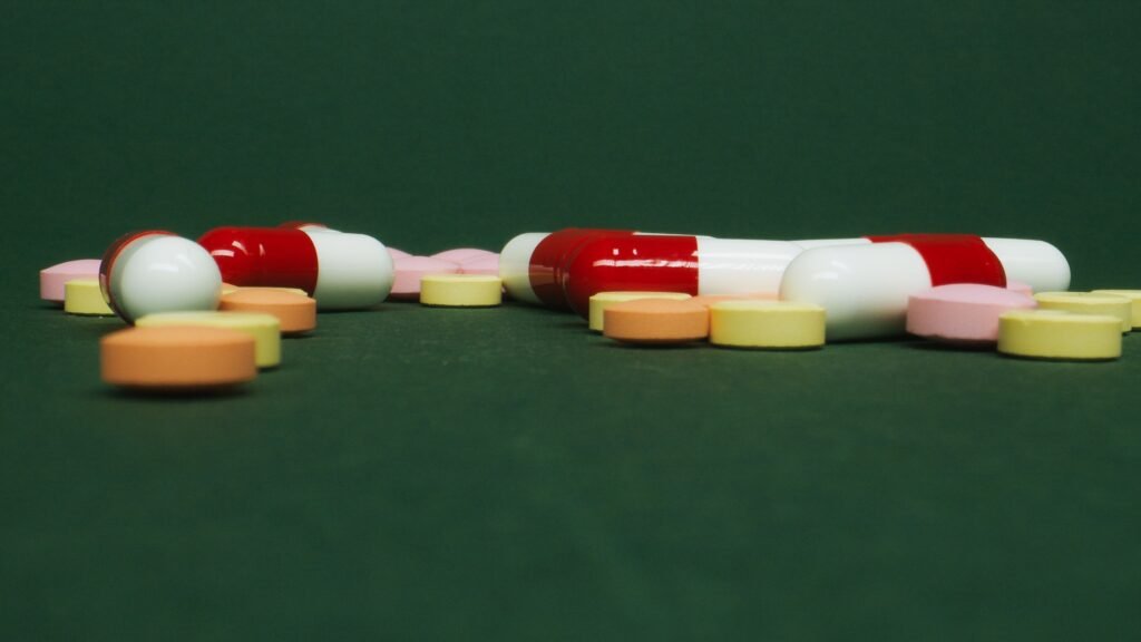
Therapeutic management of Iron Deficiency Anemia is based on three pillars: (1) identifying and treating the underlying etiology (e.g., endoscopic hemostasis, polypectomy, gluten-free diet, or hormonal control of menorrhagia), (2) pharmacological iron replacement, and (3) erythrocyte transfusion reserved exclusively for life-threatening hemodynamic instability.
1. Oral Iron Pharmacotherapy: Formulations, Mechanism, and the Alternate-Day Dosing Paradigm
Oral iron remains the first-line treatment for uncomplicated, mild-to-moderate IDA in patients with an intact, non-inflamed gastrointestinal tract.
Mechanism of Action: Oral iron salts dissolve in acidic gastric secretions to release divalent ferrous ions (Fe2+) or trivalent ferric complexes, which are absorbed across the duodenal enterocyte apical membrane via DMT1, exported into plasma via ferroportin, and delivered by transferrin to bone marrow erythroblasts to restore mitochondrial heme synthesis and globin translation. Elemental Iron is the actual amount of pure iron present in a supplement, which is the part that the human body absorbs and uses.
Pharmacological Formulations and Elemental Iron Content:
- Ferrous Sulfate (FeSO4): Contains 20% elemental iron (a standard 325 mg hydrated tablet provides 65 mg of elemental iron; a 200 mg dried tablet provides 65 mg). Most cost-effective standard of care.
- Ferrous Gluconate: Contains 12% elemental iron (a 300 to 325 mg tablet provides 35 to 38 mg of elemental iron).
- Ferrous Fumarate: Contains 33% elemental iron (a 325 mg tablet provides 106 mg of elemental iron; a 210 mg tablet provides 69 mg).
- Polysaccharide-Iron Complex and Heme Iron Polypeptide: Alternative formulations designed to improve gastrointestinal tolerability, though clinical trials demonstrate no superiority in mucosal absorption over standard ferrous salts when adjusted for elemental iron dose.
- Ferric Maltol and Sucrosomial Iron: Novel third-generation oral preparations. Ferric maltol chelates Fe3+ within a trimaltol complex that remains stable at neutral pH and delivers iron directly to the enterocyte brush border with minimal free luminal iron generation (approved for IBD-associated Iron Deficiency Anemia). Sucrosomial iron encapsulates ferric pyrophosphate within a phospholipid bilayer and sucrester matrix, enabling absorption via paracellular and transcellular M-cell pathways independent of DMT1, markedly reducing gastrointestinal adverse effects.
Dosing Strategy — The Paradigm Shift to Alternate-Day Dosing:
- Historical Dogma vs. Modern Hepcidin Kinetics: Historically, clinicians prescribed 325 mg of ferrous sulfate three times daily (195 mg elemental iron/day). However, landmark stable iron isotope pharmacokinetic studies (Stoffel et al., Lancet Haematology, Blood) demonstrated that a single oral dose of 60 mg or more of elemental iron triggers an acute spike in circulating serum hepcidin that peaks at 24 hours and remains elevated for up to 48 hours. This hepcidin surge internalizes duodenal ferroportin, reducing fractional absorption of subsequent doses taken later the same day or the following morning by 35% to 50%, while leaving unabsorbed luminal iron to cause oxidative mucosal irritation, nausea, epigastric cramping, metallic taste, and constipation via colonic microbiome shifts.
- Evidence-Based Dosing Regimen: Current AGA (2024) and BSH guidelines recommend 60 to 100 mg of elemental iron taken once daily every other day (alternate-day dosing) (e.g., 1 to 2 tablets of 325 mg ferrous sulfate on Monday, Wednesday, and Friday mornings), or a lower single daily dose of 40 to 65 mg of elemental iron once daily. Alternate-day dosing yields significantly higher fractional iron absorption (up to 30%–50% higher per milligram administered) and drastically lowers gastrointestinal discontinuation rates.
- Administration Optimization: Oral iron should be taken on an empty stomach in the morning with water or 80 to 250 mg of ascorbic acid (vitamin C) or orange juice, which chelates iron and maintains it in the reduced Fe2+ state. Patients must avoid tea, coffee, milk, calcium supplements, antacids, and fluoroquinolone/tetracycline antibiotics within 2 hours before or after dosing, and take levothyroxine at least 4 hours apart (as iron chelates thyroxine in the gut lumen).
- Administration Optimization: Oral iron should be taken on an empty stomach in the morning with water or 80 to 250 mg of ascorbic acid (vitamin C) or orange juice, which chelates iron and maintains it in the reduced Fe2+ state. Patients must avoid tea, coffee, milk, calcium supplements, antacids, and fluoroquinolone/tetracycline antibiotics within 2 hours before or after dosing, and take levothyroxine at least 4 hours apart (as iron chelates thyroxine in the gut lumen).
2. Intravenous (Parenteral) Iron Pharmacotherapy
Mechanism of Action: Intravenous iron formulations consist of a polynuclear ferric-oxyhydroxide (Fe3+) core surrounded by a stabilizing carbohydrate shell (dextran, sucrose, carboxymaltose, or derisomaltose) that prevents the toxic release of free labile iron into plasma. Following infusion, these nanoparticles are phagocytosed directly by reticuloendothelial macrophages in the spleen, liver, and bone marrow. Inside the macrophage phagolysosome, the carbohydrate shell is cleaved, and iron is either stored in ferritin or exported via ferroportin into plasma to bind transferrin, completely bypassing the duodenal mucosal bottleneck.
Clinical Indications for First-Line or Switch to IV Iron:
- Intolerance or non-adherence to oral iron due to refractory gastrointestinal side effects.
- Anatomic or mucosal malabsorption: Celiac disease, post-bariatric surgery (Roux-en-Y, sleeve gastrectomy), autoimmune atrophic gastritis, or short bowel syndrome
- Chronic Inflammatory Conditions where high systemic hepcidin blocks oral absorption: Active inflammatory bowel disease (IBD), chronic kidney disease (both non-dialysis and hemodialysis-dependent), and symptomatic heart failure with reduced ejection fraction (HFrEF).
- Ongoing Blood Loss Exceeding Oral Absorptive Capacity: Severe menorrhagia, hereditary hemorrhagic telangiectasia (Osler-Weber-Rendu disease), or refractory gastrointestinal angioectasias.
- Second and Third Trimesters of Pregnancy: When severe Iron Deficiency Anemia (Hb below 9.0–10.0 g/dL) is diagnosed late in gestation or fails to respond to oral iron, requiring rapid maternal-fetal repletion before delivery (IV iron is avoided in the first trimester due to lack of teratogenicity safety data).
Calculating Total Iron Deficit (The Ganzoni Formula):
Total cumulative parenteral iron dose ( commonly ranging from 1,000 to 1,500 mg) is classically calculated using the Ganzoni Equation:
Total Iron Deficit (mg) = Body Weight (kg) * (Target Hb – Actual Hb in g/dL) * 2.4 + Iron Stores (500 mg for adults)
Intravenous Iron Formulations and Dosing Profiles:
- Ferric Carboxymaltose (FCM / Ferinject, Injectafer): High-stability complex allowing a single high-dose infusion of 750 mg to 1,000 mg over 15 minutes (up to 1,000 mg single dose in Europe or two 750 mg doses separated by 7 days in the US). Specific Adverse Effect: Carries the highest incidence (up to 40%–70% biochemical, occasional symptomatic osteomalacia) of treatment-emergent hypophosphatemia, because intact FCM carbohydrate shells inhibit the proteolytic cleavage of fibroblast growth factor 23 (FGF23) in osteocytes, elevating intact FGF23 and inducing renal proximal tubular phosphate wasting and 1,25-dihydroxyvitamin D suppression.
- Ferric Derisomaltose (FDI / Monofer, Monoferric): Matrix structure of linear isomaltoside 1000 oligomers tightly bound to iron, permitting complete total-dose replacement of 1,000 mg (up to 20 mg/kg) in a single 20-to-30-minute infusion with a low incidence of hypophosphatemia (less than 4%).
- Low-Molecular-Weight (LMW) Iron Dextran (INFeD, CosmoFer): Can be administered as a single total-dose infusion of 1,000 mg over 1 to 4 hours following a mandatory 25 mg slow test dose over 15 minutes to monitor for hypersensitivity (note: historic high-molecular-weight iron dextran [Dexferrum], which carried high anaphylaxis risk, has been withdrawn from clinical practice).
- Iron Sucrose (Venofer): Loosely bound complex requiring smaller fractional doses of 200 mg infused over 15–30 minutes across 5 separate sessions (or 300 mg across 3–4 sessions) to reach 1,000 mg; widely used in hemodialysis centers.
- Sodium Ferric Gluconate (Ferrlecit): Administered in fractional doses of 125 mg per session over 8 dialysis sessions.
- Ferumoxytol (Feraheme): Superparamagnetic iron oxide coated with polyglucose sorbitol carboxymethyl ether, dosed at 510 mg infused over 15 minutes, repeated 3 to 8 days later (1,020 mg total). Clinical Caveat: Can transiently alter MRI signal intensity for up to 3 months post-infusion.
Managing Infusion Reactions (Fishbane Reaction vs. True Anaphylaxis): True IgE-mediated anaphylaxis to modern non-dextran IV iron occurs in fewer than 1 in 200,000 administrations. More commonly (roughly 1% of infusions), patients experience a complement-activation-related pseudo-allergy (CARPA or “Fishbane reaction”) triggered by labile free iron nanoparticles, characterized by acute self-limited facial flushing, chest tightness, and truncal/back myalgias without hypotension, wheezing, or periorbital edema. Management consists of pausing the infusion for 15 minutes until symptoms subside and restarting at 50% of the initial infusion rate; administering empirical diphenhydramine (an H1-antihistamine) is contraindicated for mild Fishbane reactions as it causes sedation, tachycardia, and hypotension that mimic worsening anaphylaxis.
3. Duration of Therapy and Monitoring Response
Kinetics of Hematologic Recovery:
- 24 to 72 Hours: Subjective improvement in pagophagia, fatigue, and intracellular enzyme activity.
- Days 4 to 10 (Peak at Days 7–10): Reticulocytosis occurs in the peripheral blood, accompanied by an immediate surge in reticulocyte hemoglobin content (CHr).
- Weeks 2 to 4: Hemoglobin concentration should increase by at least 1.0 to 2.0 g/dL (10 to 20 g/L) within 2 to 4 weeks of initiating oral or IV iron. Failure to achieve a 1.0 g/dL Hb rise at 4 weeks defines oral iron refractoriness and necessitates checking adherence, ongoing occult bleeding, celiac disease, H. pylori, or switching to IV iron.
- Weeks 6 to 8: Complete normalization of hemoglobin and MCV.
Total Duration of Oral Treatment: Crucially, oral iron therapy must not be discontinued when hemoglobin normalizes. Therapy must be continued for an additional 3 to 6 months after hemoglobin normalization (until serum ferritin exceeds 50 to 100 ng/mL) to completely replenish reticuloendothelial macrophage and hepatic storage iron pools and prevent rapid relapse.
- Monitoring After IV Iron: Serum ferritin should not be measured within the first 4 weeks following an IV iron infusion, as circulating uncleared carbohydrate-iron complexes falsely elevate the ferritin assay; repeat Complete Blood Count and iron panel assessment is optimally performed at 4 to 8 weeks post-infusion.
4. Red Blood Cell Transfusion Criteria
Because chronic Iron Deficiency Anemia allows gradual physiological compensation—specifically increased erythrocyte 2,3-bisphosphoglycerate (2,3-BPG) synthesis, which shifts the oxyhemoglobin dissociation curve to the right (P50 increases from 27 mmHg to over 30 mmHg) to enhance tissue oxygen unloading—even profoundly anemic patients (Hb 5.0–6.5 g/dL) are often hemodynamically stable at rest. Per AABB guidelines, packed red blood cell (pRBC) transfusion is restricted to:
- Hemodynamic instability or active, brisk hemorrhage.
- Hemoglobin below 7.0 g/dL in hospitalized patients with acute symptomatic cardiopulmonary compromise, or below 8.0 g/dL in patients with active coronary artery disease, acute coronary syndrome, or heart failure.
- When transfused in chronic severe Iron Deficiency Anemia, single units should be administered slowly (one unit at a time with clinical reassessment) to avoid precipitating transfusion-associated circulatory overload (TACO) in a hypervolemic, compensated vasculature.
9. Iron Deficiency Anemia Prevention and Outlook, Prognosis, and Chronic Clinical Outcome
Section Summary:
The long-term prognosis of iron deficiency anemia is excellent when the underlying cause is benign and iron stores are fully replenished, with complete resolution of hematologic and epithelial abnormalities. However, untreated chronic iron deficiency in infancy can cause irreversible neurocognitive and psychomotor deficits, while severe maternal anemia increases the risk of preterm birth, low birth weight, and postpartum hemorrhage.
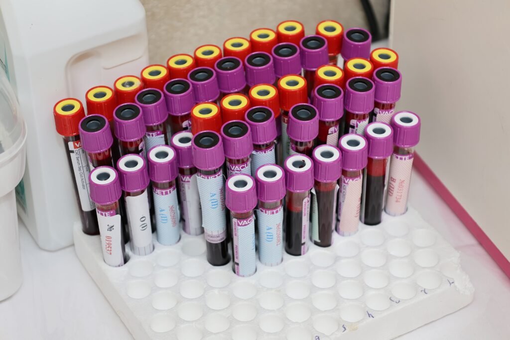
Primary and Secondary Prevention Strategies
- Dietary Optimization and Food Fortification: Consuming bioavailable heme iron sources (lean red meat, poultry, fish, shellfish) alongside non-heme plant sources (lentils, chickpeas, spinach, pumpkin seeds, fortified cereals) paired with vitamin C-rich foods (citrus fruits, bell peppers, broccoli) to enhance duodenal reduction of Fe3+, while separating tea, coffee, and calcium intake from iron-rich meals. At a population level, WHO recommends universal iron fortification of wheat and maize flour in endemic areas.
- Perinatal and Obstetrical Prophylaxis: Routine low-dose oral iron supplementation (30 to 60 mg elemental iron daily or intermittent weekly dosing) during pregnancy, coupled with delayed umbilical cord clamping (for 60 to 180 seconds after birth), which transfers an additional 80 to 100 mL of placental blood to the neonate and boosts infant iron stores for the first 6 months of life.
- Pediatric Prophylaxis: The American Academy of Pediatrics recommends prophylactic oral iron drops (2 mg/kg/day starting at 1 month of age) for preterm infants, and 1 mg/kg/day starting at 4 months of age for exclusively breastfed term infants until iron-rich complementary foods are established at 6 months, alongside strict avoidance of unmodified cow’s milk before 12 months of age.
- Blood Donor Protection: Monitoring donor serum ferritin and recommending a 60-day course of low-dose oral iron (18 to 38 mg elemental iron daily) following whole-blood donation.
Prognosis and Chronic Clinical Outcomes
- Adults: When the primary etiology is identified and corrected (e.g., eradication of H. pylori, initiation of a gluten-free diet in celiac disease, or management of uterine fibroids) and iron stores are replenished, the hematologic prognosis is curative. Mucosal signs such as glossitis and koilonychia resolve completely over 3 to 6 months as epithelial and nail matrices regenerate. Conversely, if Iron Deficiency Anemia in an older adult is secondary to an undiagnosed gastrointestinal malignancy, overall survival is dictated by the oncologic stage at diagnosis—underscoring why timely endoscopic evaluation is life-saving.
- Neurodevelopmental Sequelae in Infants and Children: Iron is essential during the critical neurodevelopmental window of the first 1,000 days of life (from conception to age 2 years) for oligodendrocyte myelination, hippocampal dendritogenesis, and monoaminergic striatal circuitry. Longitudinal cohort studies demonstrate that severe, prolonged Iron Deficiency Anemia during infancy is associated with persistent deficits in auditory and visual evoked potentials, motor coordination, executive function, and arithmetic/reading scores that may remain partially irreversible into adolescence even after subsequent iron normalization.
- Maternal and Fetal Outcomes in Pregnancy: Untreated moderate-to-severe maternal Iron Deficiency Anemia during pregnancy increases the relative risk of preterm delivery (by 1.5- to 2-fold), intrauterine growth restriction (IUGR), low birth weight, neonatal iron store depletion, and maternal cardiovascular decompensation or death during peripartum hemorrhage, as well as postpartum depression and impaired maternal-infant bonding.
10. Iron Deficiency Anemia Interconnection with Other Medical Conditions
Section Summary:
Iron deficiency anemia intersects bidirectionally with major systemic pathologies across cardiology, nephrology, gastroenterology, and endocrinology. In congestive heart failure and chronic kidney disease, functional and absolute iron deficiency independently increase hospitalization and mortality rates regardless of hemoglobin levels. Additionally, iron deficiency alters platelet reactivity, accelerates hypoxic pulmonary vasoconstriction, and distorts glycated hemoglobin (HbA1c) measurements in diabetes mellitus.

1. Heart Failure (HFrEF and HFpEF) and Cardiorenal Syndrome
Iron deficiency is present in approximately 50% of patients with chronic heart failure and up to 80% of those hospitalized with acute decompensated heart failure, frequently occurring even in the absence of anemia.
- Pathophysiological Link: Splanchnic venous congestion causes bowel wall edema (impairing duodenal iron absorption), concomitant antiplatelet/anticoagulant therapy causes occult GI blood loss, and chronic low-grade inflammatory cytokines (IL-6, TNF-α) elevate hepcidin to trap iron within macrophages. Crucially, cardiomyocytes possess the highest mitochondrial density of any human cell; myocardial iron depletion directly reduces Complex I, II, and III iron-sulfur cluster activity and ATP generation, impairing left ventricular contractility and lusitropy.
- Clinical Trial Evidence and Guidelines: Landmark randomized controlled trials (FAIR-HF, CONFIRM-HF, AFFIRM-AHF, IRONMAN) demonstrated that intravenous iron replacement (specifically with ferric carboxymaltose or ferric derisomaltose) in HFrEF patients with iron deficiency (defined as ferritin below 100 ng/mL, or ferritin 100–299 ng/mL with TSAT below 20%) significantly improves 6-minute walk distance, New York Heart Association (NYHA) functional class, quality of life, and reduces recurrent hospitalizations for heart failure—even in patients whose baseline hemoglobin is completely normal. Consequently, European Society of Cardiology (ESC) and AHA/ACC/HFSA guidelines assign a Class I/IIa recommendation for routine screening and IV iron repletion in HFrEF.
2. Chronic Kidney Disease (CKD) and Renal Anemia
In Chronic Kidney Disease stages 3 through 5, anemia stems from a dual defect: reduced erythropoietin synthesis by fibrotic renal peritubular interstitial cells and profound absolute and functional iron deficiency.
- Pathophysiological Link: Because hepcidin (a 2.7 kDa peptide) is normally cleared via glomerular filtration and tubular degradation, declining glomerular filtration rate (GFR) coupled with uremic inflammation causes plasma hepcidin levels to rise 5- to 10-fold, locking iron inside macrophages and blocking intestinal absorption. Furthermore, initiating Erythropoiesis-Stimulating Agents (ESAs) without adequate iron repletion induces functional iron deficiency and increases the risk of ESA-induced hypertension, stroke, and vascular access thrombosis (partially driven by iron-deficiency-induced reactive thrombocytosis).
- KDIGO Guideline Thresholds: The Kidney Disease: Improving Global Outcomes (KDIGO) guidelines recommend initiating iron therapy (preferentially IV iron in hemodialysis patients, as validated by the PIVOTAL trial, which showed high-dose proactive IV iron sucrose reduced cardiovascular events and ESA dose requirements) when TSAT is 30% or below and serum ferritin is 500 ng/mL or below.
3. Type 2 Diabetes Mellitus and Spurious HbA1c Elevation
Iron deficiency anemia exerts a profound pre-analytical artifact on the diagnosis and monitoring of Type 2 Diabetes Mellitus (T2DM):
- False Elevation of HbA1c in Untreated Iron Deficiency Anemia: Untreated iron deficiency anemia falsely elevates HbA1c concentrations by 0.5% to 1.5% independent of true ambient glycemia. Two mechanisms account for this phenomenon: (1) reduced bone marrow erythropoiesis slightly prolongs the average circulating age of surviving erythrocytes, extending their exposure time to non-enzymatic glycation, and (2) heightened oxidative stress (elevated malondialdehyde) in iron-deficient erythrocytes accelerates the irreversible ketoamine linkage of glucose to the N-terminal valine of the β-globin chain. Reliance on HbA1c alone in an undiagnosed iron-deficient patient can lead to a false diagnosis of prediabetes or diabetes, or dangerous over-intensification of hypoglycemic therapy.
- False Lowering of HbA1c During Iron Replacement Therapy: Conversely, during the first 4 to 8 weeks after initiating oral or intravenous iron therapy, the rapid flood of young, unglycated reticulocytes into the circulation falsely lowers the HbA1c value. Clinicians should monitor glycemic control via fasting plasma glucose or continuous glucose monitoring (CGM) until iron stores and red cell turnover stabilize.
4. Thromboembolic Disease, Stroke, and Pulmonary Hypertension
- Venous Thromboembolism (DVT and Pulmonary Embolism) and Ischemic Stroke: Severe Iron Deficiency Anemia is an independent, under-recognized risk factor for both arterial and venous thrombosis (including cerebral venous sinus thrombosis, deep vein thrombosis, and pulmonary embolism). The prothrombotic state is mediated by reactive thrombocytosis, increased erythropoietin-driven platelet P-selectin expression and hyper-aggregability, and turbulent high-velocity blood flow coupled with inflexible microcytic erythrocytes that displace platelets toward the endothelial wall, increasing endothelial shear stress.
- Pulmonary Arterial Hypertension (PAH): Iron is a mandatory cofactor for prolyl hydroxylases (PHDs) that degrade HIF-1α and HIF-2α in the pulmonary vasculature. Iron deficiency stabilizes pulmonary endothelial HIF-2α even under normoxic conditions, upregulating endothelin-1 and exaggerating hypoxic pulmonary vasoconstriction and pulmonary vascular remodeling.
11. Iron Deficiency Anemia Frequently Asked Questions (FAQs)
Section Summary:
Patients frequently seek clarity regarding the distinction between low ferritin and clinical anemia, why oral supplements cause gastrointestinal distress, how diet compares to pharmacological replacement, and why endoscopic procedures are required even without abdominal symptoms. Addressing these questions with evidence-based physiological explanations improves therapeutic adherence and ensures timely completion of diagnostic evaluations.

1. Can I have symptoms of iron deficiency if my hemoglobin is completely normal?
Yes. This clinical entity is known as non-anemic iron deficiency (NAID) or Stage I/II iron depletion. Because the bone marrow prioritizes erythropoiesis above all other tissues, the body strips iron from skeletal muscle myoglobin, hair follicles, and central nervous system enzymes (such as tyrosine hydroxylase and cytochrome c oxidase) long before hemoglobin drops below normal. Patients with a serum ferritin below 30 ng/mL and a normal hemoglobin frequently experience profound fatigue, brain fog, restless legs syndrome, hair shedding, and exercise intolerance, all of which improve significantly with iron repletion.
2. Why should I take my iron pill every other day instead of three times a day?
When you take a standard dose of oral iron (60 mg or more of elemental iron), your liver responds within hours by releasing a surge of the hormone hepcidin, which acts as a molecular lock on the iron-exporting gates (ferroportin) of your intestinal cells for the next 24 hours. If you take another pill that evening or the next morning, your gut blocks most of it from entering your bloodstream; the unabsorbed iron simply sits in your intestines, causing nausea, cramps, and constipation. Waiting 48 hours between doses allows hepcidin to drop back to baseline, resulting in higher iron absorption and far fewer side effects.
3. Why do my stools turn black when I take oral iron, and does it mean I am bleeding?
Unabsorbed dietary and supplemental iron reacts with hydrogen sulfide produced by normal bacteria in the colon to form iron sulfide, a completely harmless compound that turns stool a dark greenish-black color. While dark stools are an expected sign that you are taking your supplement (and do not trigger a false-positive result on modern fecal immunochemical tests [FIT]), true upper gastrointestinal bleeding causes foul-smelling, tarry stools (melena). If black stools are accompanied by dizziness, vomiting, or worsening anemia, prompt medical evaluation is required.
4. Can I cure already-established Iron Deficiency Anemia through diet alone (such as eating spinach and red meat)?
No. Once frank Iron Deficiency Anemia has developed, total body iron stores are depleted by roughly 1,000 to 1,500 mg. Even an optimal iron-rich diet provides only about 15 to 20 mg of dietary iron per day, of which the duodenum can absorb at most 2 to 4 mg/day. Relying on diet alone would take well over a year to rebuild your hemoglobin and ferritin reserves, assuming zero ongoing blood loss. Dietary optimization is vital for long-term prevention, but therapeutic pharmacological supplementation (oral or IV) is mandatory to treat established Iron Deficiency Anemia.
5. Why does my doctor insist on a colonoscopy and upper endoscopy if I have no stomach pain or visible blood in my stool?
In adult men and postmenopausal women, there is no physiological pathway for significant iron loss. The most common cause of newly diagnosed IDA in these groups is slow, microscopic (“occult”) bleeding from the gastrointestinal tract—and in 8% to 15% of cases, the source is an early-stage colorectal or gastric cancer or precancerous polyp that leaks only a few drops of blood per day (completely invisible to the naked eye and painless). Performing bidirectional endoscopy catches these lesions at an early, curable stage.
12. Questions to Ask the Doctor About Iron Deficiency Anemia
Section Summary:
Empowering patients with targeted, clinically precise questions transforms routine medical consultations into collaborative diagnostic partnerships. Key inquiries should focus on clarifying the exact serum ferritin and transferrin saturation values, identifying the root anatomical cause of iron loss, optimizing oral dosing schedules to minimize side effects, establishing criteria for intravenous infusions, and scheduling follow-up laboratory monitoring.

- “What are my exact hemoglobin, MCV, serum ferritin, and transferrin saturation (TSAT) numbers, and what is our specific target ferritin level before I stop treatment?” (Ensures treatment continues until storage pools—not just hemoglobin—are fully restored above 50 to 100 ng/mL).
- “Now that we know I have Iron Deficiency Anemia, what diagnostic tests—such as celiac serology, H. pylori testing, or upper and lower endoscopy—do I need to find the root cause of my iron loss?” (Prevents treating the lab value with iron pills while missing an underlying malabsorptive or bleeding condition).
- “Would I benefit from an alternate-day (every-other-day) oral iron dosing schedule with vitamin C to maximize absorption and minimize stomach upset?” (Aligns the prescription with modern hepcidin-based pharmacokinetic guidelines).
- “Do any of my current daily medications—such as proton pump inhibitors (like omeprazole), calcium supplements, or thyroid hormone (levothyroxine)—interfere with how my body absorbs iron, and how should I space them out?” (Prevents luminal chelation and malabsorption).
- “Given my medical history or the severity of my deficiency, am I a candidate for a single-visit intravenous iron infusion instead of months of oral tablets?” (Especially relevant for patients with Inflammatory Bowel Disease, heart failure, Chronic Kidney Disease, heavy menstrual bleeding, or prior bariatric surgery).
- “When should we repeat my complete blood count and reticulocyte or ferritin panel to confirm that my bone marrow is responding to treatment?” (Establishes the critical 2-to-4-week check for a 1.0 – 2.0 g/dL Hb rise and the 3-month storage recheck).
13. Internal Link Section: Clinical and Pharmacological Interconnections
Section Summary:
Iron deficiency anemia rarely exists in clinical isolation; it actively modifies the pathophysiology, diagnostic interpretation, and pharmacological risks of cardiovascular, renal, metabolic, and thromboembolic disorders. Exploring the evidence-based clinical guides below illuminates how systemic iron homeostasis intersects with diagnostic blood panels, heart failure, coronary ischemia, chronic kidney disease, diabetes mellitus, and pulmonary embolism.
To build a complete clinical picture of how Iron Deficiency Anemia interacts with systemic organ systems and cardiovascular pharmacotherapy, explore UncutMed’s interconnected clinical monographs:
- [Complete Guide to Blood Tests: Interpreting Your CBC and Metabolic Panels]
Clinical & Diagnostic Connection: Learn how to systematically interpret the Complete Blood Count (CBC)—including Hemoglobin, Hematocrit, MCV, MCH, MCHC, and RDW—alongside the Mentzer Index and iron studies to differentiate Iron Deficiency Anemia from thalassemia and anemia of chronic inflammation.
- [Congestive Heart Failure (CHF): Pathophysiology, Stages, and Guideline-Directed Medical Therapy]
Clinical & Pharmacological Connection: Up to 50% of patients with Heart Failure have comorbid iron deficiency, which starves cardiac myocytes of mitochondrial ATP. Discover why major cardiology guidelines mandate screening for ferritin (below 100 ng/mL) and TSAT (below 20%) and administering intravenous ferric carboxymaltose or ferric derisomaltose to reduce heart failure hospitalizations
- [Chronic Kidney Disease (CKD): Pathophysiology, Staging, and Clinical Management]
Clinical & Pharmacological Connection: Explore how declining glomerular filtration rate impairs renal erythropoietin (EPO) production while preventing the clearance of hepcidin, creating a dual block of absolute and functional iron deficiency that must be corrected before initiating Erythropoiesis-Stimulating Agents (ESAs).
- [Coronary Artery Disease (CAD): Pathophysiology, Diagnosis, and Management]
Clinical & Pharmacological Connection: In patients with atherosclerotic coronary stenosis, IDA-induced reductions in arterial oxygen content (CaO2) accompanied by compensatory reflex tachycardia simultaneously decrease myocardial oxygen supply and increase myocardial oxygen demand, precipitating Type 2 myocardial infarction or unstable angina. Furthermore, Dual Antiplatelet Therapy (DAPT) with aspirin and P2Y12 inhibitors (clopidogrel, ticagrelor) alongside proton pump inhibitors is a leading iatrogenic driver of occult GI bleeding and iron malabsorption.
- [Atrial Fibrillation (AFib): Mechanisms, Stroke Risk, and Anticoagulation]
Clinical & Pharmacological Connection: Hyperdynamic states and myocardial hypoxia from severe IDA can trigger rapid ventricular response and paroxysmal atrial fibrillation. Crucially, the development of new-onset Iron Deficiency Anemia in a patient taking Direct Oral Anticoagulants (DOACs) or warfarin for AFib is a clinical red flag that frequently unmasks an underlying gastrointestinal malignancy or angiodysplasia.
- [Type 2 Diabetes Mellitus (T2DM): Pathophysiology, Diagnostic Criteria, and Pharmacology]
Clinical & Diagnostic Connection: Understand how untreated Iron Deficiency Anemia falsely elevates HbA1c by prolonging erythrocyte survival and increasing lipid peroxidation, whereas active iron replacement therapy transiently falsely lowers HbA1c due to reticulocytosis—critical nuances when titrating SGLT2 inhibitors (which themselves stimulate erythropoiesis and drop hepcidin) or insulin.
- [Pulmonary Embolism (PE): Pathophysiology, Risk Stratification, and Management]
Clinical & Hematologic Connection: Severe Iron Deficiency Anemia induces reactive thrombocytosis, increases platelet P-selectin expression, and elevates turbulent endothelial shear stress, acting as a potent prothrombotic catalyst for venous thromboembolism (VTE) and Pulmonary Embolism. Conversely, anticoagulant therapy for PE can precipitate heavy menstrual or gastrointestinal bleeding, worsening IDA.
- [Hypertension: Pathophysiology, Secondary Causes, and Pharmacological Treatment]
Clinical & Hemodynamic Connection: Chronic severe anemia triggers nitric oxide-mediated peripheral vasodilation, reduced blood viscosity, and a widened pulse pressure with elevated systolic blood pressure, while iron deficiency in the pulmonary vasculature stabilizes HIF-2α to drive pulmonary hypertension.
- [Atherosclerosis: Endothelial Dysfunction, Plaque Biology, and Lipid Management]
Clinical & Molecular Connection: Examine the delicate balance of iron in vascular biology—where iron deficiency impairs antioxidant enzymes (catalase and glutathione peroxidase), while excess free labile iron catalyzes Fenton-reaction oxidation of LDL cholesterol inside subendothelial macrophages (foam cells).
14. High-Trust Peer-Reviewed References, Sources, and Clinical Guidelines
Section Summary:
This clinical monograph is synthesized from current international practice guidelines, landmark randomized controlled trials, and authoritative medical textbooks. Primary references include consensus directives from the World Health Organization, the American Gastroenterological Association, the British Society for Haematology, the European Society of Cardiology, and foundational chapters from Harrison’s Principles of Internal Medicine and Williams Hematology.
International Clinical Practice Guidelines
- World Health Organization (WHO). Guideline on haemoglobin cutoffs to define anaemia in individuals and populations. Geneva: World Health Organization; 2024. ISBN: 978-92-4-008854-2.
- World Health Organization (WHO). WHO guideline on use of ferritin concentrations to assess iron status in individuals and populations. Geneva: World Health Organization; 2020. ISBN: 978-92-4-000296-8.
- American Gastroenterological Association (AGA). DeLoughery TG, Jackson CS, Ko CW, Rockey DC. AGA Clinical Practice Update on Management of Iron Deficiency Anemia: Expert Review. Clinical Gastroenterology and Hepatology. 2024;22(8):1575–1583.
- American Gastroenterological Association (AGA). Ko CW, Siddique SM, Patel A, et al. AGA Clinical Practice Guidelines on the Gastrointestinal Evaluation of Iron Deficiency Anemia. Gastroenterology. 2020;159(3):1085–1094.
- British Society for Haematology (BSH). Fletcher A, Forbes A, Svenson N, Wayne Thomas D. Guideline for the laboratory diagnosis of iron deficiency in adults (excluding pregnancy and children). British Journal of Haematology. 2022;196(3):523–529.
- British Society of Gastroenterology (BSG). Snook J, Bhala N, Beales ILP, et al. British Society of Gastroenterology guidelines for the management of iron deficiency anaemia in adults. Gut. 2021;70(11):2030–2051.
- British Society for Haematology (BSH). Pavord S, Daru J, Prasannan N, et al. UK guidelines on the management of iron deficiency in pregnancy. British Journal of Haematology. 2020;188(6):819–830.
- European Society of Cardiology (ESC). McDonagh TA, Metra M, Adamo M, et al. 2023 Focused Update of the 2021 ESC Guidelines for the diagnosis and treatment of acute and chronic heart failure. European Heart Journal. 2023;44(37):3627–3639.
- Kidney Disease: Improving Global Outcomes (KDIGO). Babitt JL, Eisenga MF, Haase VH, et al. Controversies in optimal anemia management: conclusions from a Kidney Disease: Improving Global Outcomes (KDIGO) Conference. Kidney International. 2021;99(6):1280–1295.
- National Institutes of Health (NIH) Office of Dietary Supplements. Iron: Fact Sheet for Health Professionals. U.S. Department of Health and Human Services; Updated 2024.
Authoritative Medical Textbooks
- Camaschella C,Weiss G. “Iron-Deficiency Anemia and Megaloblastic Anemias.” In: Loscalzo J, Fauci A, Kasper D, Hauser S, Longo D, Jameson JL, eds. Harrison’s Principles of Internal Medicine. 21st ed. New York, NY: McGraw-Hill Education; 2022:chap 97.
- Ganz T, Nemeth E. “Iron Metabolism and Iron Deficiency Anemia.” In: Kaushansky K, Prchal JT, Burns LJ, Lichtman MA, Levi M, Linch DC, eds. Williams 2021: chap.Hematology. 10th ed. New York, NY: McGraw-Hill Education; 2021: chap 43.
- Andrews NC, Fleming MD. “Pathobiology of the Human Enterocyte and Systemic Iron Homeostasis.” In: Hoffman R, Benz EJ, Silberstein LE, Heslop HE, Weitz JI, Salama ME, Abutalib SA, eds. Hematology: Basic Principles and Practice. 8th ed. Philadelphia, PA: Elsevier; 2023:chap 36.
- Aster JC, Abbas AK. “Red Blood Cell and Bleeding Disorders.” In: Kumar V, Abbas AK, Aster JC, eds. Robbins & Cotran Pathologic Basis of Disease. 10th ed. Philadelphia, PA: Elsevier; 2021:chap 14.
Landmark Peer-Reviewed Journal Articles & Reviews
- Camaschella C. Iron-Deficiency Anemia. New England Journal of Medicine. 2015;372(19):1832–1843.
- Pasricha SR, Tye-Din J, Muckenthaler MU, Swinkels DW. Iron deficiency. The Lancet. 2021;397(10270):233–248.
- Stoffel NU, Cercamondi CI, Brittenham G, et al. Iron absorption from oral iron supplements given on consecutive versus alternate days and as single morning doses versus twice-daily split dosing in iron-depleted women: two open-label, randomised controlled trials. The Lancet Haematology. 2017;4(11):e524–e533.
- Stoffel NU, Zeder C, Brittenham GM, Moretti D, Zimmermann MB. Iron absorption from supplements is greater with alternate day than with consecutive day dosing in iron-deficient anemic women. Haematologica. 2020;105(5):1232–1239.
- Ganz T, Nemeth E. Iron homeostasis in host defence and inflammation. Nature Reviews Immunology. 2015;15(8):500–510.
- Kautz L, Jung G, Valore EV, Rivella S, Nemeth E, Ganz T. Identification of erythroferrone as an erythroid regulator of iron metabolism. Nature Genetics. 2014;46(7):678–684.
- Wolf M, Rubin J, Achebe M, et al. Effects of Iron Isomaltoside vs Ferric Carboxymaltose on Hypophosphatemia in Iron-Deficiency Anemia: Two Randomized Clinical Trials. JAMA. 2020;323(5):432–443.
Featured Image: 3D medical illustration of normal Red Blood Cells vs. iron-depleted red blood cells inside the lumen of a blood vessel. The image was created with the help of artificial intelligence.
© UncutMed — Medical knowledge, precise, truthful, unfiltered. The current article was last modified on October 24, 2026. This article was clinically authored and reviewed under international evidence-based standards. Always consult your personal healthcare physician before modifying any medical therapy.