© UncutMed — Medical knowledge, precise, truthful, unfiltered. The current article was last modified on October 7, 2026. This article was clinically authored and reviewed under international evidence-based standards. Always consult your personal healthcare physician before modifying any medical therapy.
Medical Disclaimer: This article is for informational and educational purposes only. It does not constitute medical advice, formal diagnosis, or treatment recommendations. Always consult a qualified healthcare professional regarding any metabolic condition or therapeutic plan.
Atherosclerosis is the primary pathological substrate underlying coronary artery disease, ischemic cerebrovascular disease, and peripheral arterial disease. Despite its status as the leading contributor to global cardiovascular mortality, public and clinical discourse frequently reduces this intricate immunometabolic disease to an inaccurate mechanical analogy: grease accumulating on the inside of a household pipe.
This guide dismantles that plumbing myth. Atherosclerosis (arterial plaque) is not a disease of luminal fouling; it is a chronic, non-resolving inflammatory and fibroproliferative disorder of the tunica intima—the inner structural layer of the arterial wall. This clinical reference details the cellular mechanisms, hemodynamic forces, phenotypic transitions, and clinical endpoints that define arterial plaque.
1. Arteriosclerosis (Arterial Plaque) Medical Definition and Vascular Architecture
Section Summary:
Atherosclerosis is an intramural, fibro-inflammatory disease of large and medium-sized arteries occurring within the tunica intima, rather than a superficial accumulation of debris in the lumen. It is pathologically distinct from arteriosclerosis, which describes non-atheromatous arterial stiffening. Understanding its intramural architecture explains why vascular plaque cannot be scrubbed away and why it permanently remodels the vessel wall.
The Anatomical Reality of Arterial Plaque: Intramural, Not Luminal
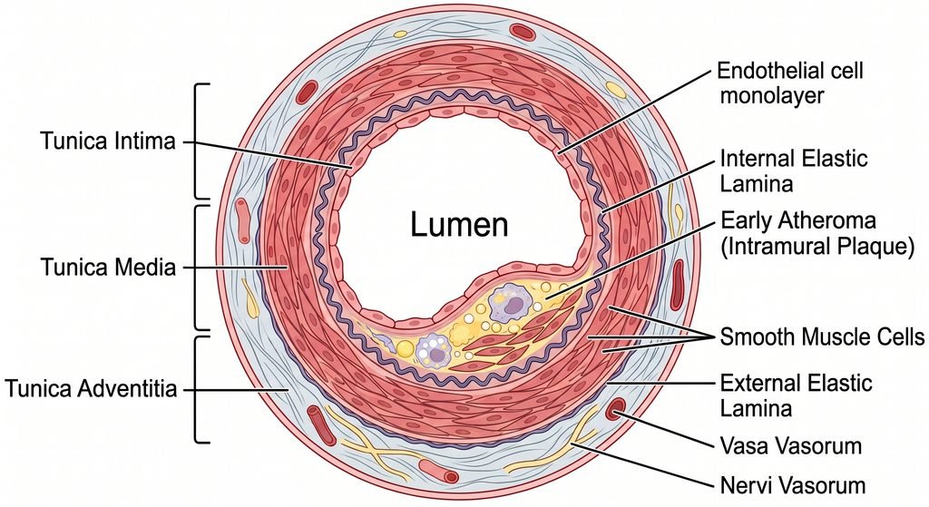
Healthy large and medium-sized arteries consist of three distinct concentric tunics:
- Tunica Intima: The innermost layer, bounded luminally by a continuous monolayer of vascular endothelial cells anchored to a basement membrane, resting on an extracellular matrix of proteoglycans and collagen. It terminates peripherally at the internal elastic lamina.
- Tunica Media: The middle structural layer, composed of concentric layers of contractile vascular smooth muscle cells (VSMCs) interspersed with elastic lamellae and collagen. It is bounded externally by the external elastic lamina.
- Tunica Adventitia: The outermost connective tissue sheath, comprising fibroblasts, fibrillar collagen, perivascular nerves, and the vasa vasorum (the microvascular network that supplies blood to the outer media of thick-walled arteries).
Atherosclerosis (arterial plaque) is an intramural disease. Pathological lipid accumulation, immune infiltration, cell death, and matrix remodeling occur entirely beneath the endothelial monolayer, inside the extracellular matrix of the tunica intima.
The circulating blood does not directly contact the atheroma during development. Instead, the lesion expands within the arterial wall itself, pushing the endothelial surface inward toward the lumen or outward into the perivascular space. Arterial Plaque is not a superficial crust that can be physically scrubbed or chemically flushed away; it is a permanent structural remodeling of the artery wall.
Classifying Arterial Pathologies: Terminological Precision
Clinical literature frequently conflates several distinct vascular conditions that may sound similar in the name, but are fundamentally different from a pathophysiological perspective. Precise differentiation is essential for diagnostic accuracy:
| Pathological Entity | Primary Anatomical Site | Primary Histopathological Feature | Typical Clinical Consequences |
| Atherosclerosis | Large and medium-sized elastic and muscular arteries (e.g., coronary, carotid, iliac, aorta) | Intimal fibro-fatty plaques consisting of a lipid/necrotic core and a collagenous fibrous cap | Acute coronary syndromes, ischemic stroke, peripheral arterial disease |
| Arteriosclerosis | Systemic arterial tree (large conduit arteries) | Generalized loss of elastin fibers, cross-linking of collagen, and structural stiffening of arterial walls | Isolated systolic hypertension, increased pulse pressure, left ventricular hypertrophy |
| Hyaline Arteriolosclerosis | Arterioles (small resistance vessels, diameter < 100 µm) | Homogeneous pink hyaline thickening of the arteriolar wall with luminal narrowing caused by extravasated plasma proteins and basement membrane duplication | Glomerulosclerosis, lacunar brain infarcts, nephrosclerosis (driven by chronic hypertension and diabetes) |
| Hyperplastic Arteriolosclerosis | Arterioles | Concentric, laminated, “onion-skin” thickening of arteriolar walls with smooth muscle hyperplasia and duplicated basement membranes | Malignant hypertension, acute renal failure (flea-bitten kidney) |
| Mönckeberg Medial Sclerosis | Tunica media of medium and small muscular arteries (often tibial, radial) | Dystrophic calcification within the internal elastic lamina and tunica media without intimal inflammation or luminal stenosis | Elevated ankle-brachial index (falsely high), vascular non-compressibility; minimal tissue ischemia |
2. Atherosclerosis (Arterial Plaque) In-Depth Molecular and Cellular Pathophysiology
Section Summary:
The pathophysiology of atherosclerosis progresses through distinct cellular phases initiated by disturbed endothelial shear stress and subendothelial ApoB particle retention. Subsequent lipoprotein oxidation triggers monocyte recruitment, foam cell formation via scavenger receptors, and NLRP3 inflammasome activation. Defective efferocytosis leads to necrotic core development, while vascular smooth muscle phenotypic switching produces a collagenous fibrous cap vulnerable to enzymatic cleavage by matrix metalloproteinases.
The pathobiology of atherosclerosis spans decades. It progresses through a defined biological sequence: hemodynamic injury, subendothelial particle trapping, immune activation, non-resolving inflammation, cell death, and structural matrix remodeling.
Step 1: Hemodynamic Shear Stress and Endothelial Glycocalyx Disruption
The healthy vascular endothelium is lined by the endothelial glycocalyx, a brush-like network of proteoglycans, glycosaminoglycans (heparan sulfate, chondroitin sulfate), and adsorbed plasma proteins. The glycocalyx serves as a physical barrier and a primary mechanotransducer.
In straight segments of the vascular tree, blood flow is uniform and laminar. This generates high steady laminar shear stress (> 15 dynes/cm²), which activates mechanosensors that upregulate Krüppel-like Factor 2 (KLF2) and Endothelial Nitric Oxide Synthase (eNOS). Nitric oxide (NO) diffuses into the underlying vascular smooth muscle to maintain vasodilation, inhibits leukocyte adhesion molecules, prevents platelet aggregation, and suppresses smooth muscle proliferation.
At arterial bifurcations, inner curves, and branch ostia (e.g., the carotid bulb, coronary bifurcations, and the infrarenal abdominal aorta), fluid dynamics change. Flow becomes turbulent, creating low, oscillatory shear stress (disturbed flow with directional oscillation and shear < 4 dynes/cm²).
Disturbed flow degrades the endothelial glycocalyx, downregulates KLF2, downregulates eNOS, and activates the transcription factor Nuclear Factor kappa-B (NF-κB). This transformation converts the endothelium from a quiescent, anti-thrombotic surface into an active, permeable, pro-adhesive, and pro-inflammatory phenotype.
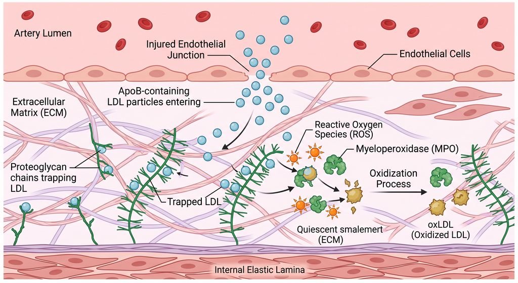
Step 2: The Response-to-Retention Hypothesis and Proteoglycan Trapping
Circulating lipoproteins do NOT trigger plaque formation simply by flowing past the vessel wall; they must cross the endothelial barrier and become immobilized.
Atherosclerosis requires the retention of Apolipoprotein B-100 (ApoB)-containing particles (including low-density lipoproteins [LDL], very-low-density lipoproteins [VLDL], intermediate-density lipoproteins [IDL], and Lipoprotein(a) [Lp(a)]). Under low shear stress and endothelial permeability:
- ApoB lipoproteins enter the subendothelial intimal space via transcytosis (mediated by scavenger receptor class B type 1 [SR-B1] and the activin-like kinase 1 [ALK1] pathway).
- Once inside the intima, basic amino acid residues (positively charged lysine and arginine) on the ApoB-100 protein interface with negatively charged sulfate groups on intimal extracellular matrix proteoglycans—specifically versican, biglycan, and decorin.
- This electrostatic interaction traps the lipoprotein particles within the intima, prolonging their residence time from hours to weeks and months.
Step 3: Subendothelial Lipoprotein Oxidation and Modification
Trapped lipoproteins are isolated from circulating plasma antioxidants like alpha-tocopherol and ascorbic acid. The trapped particles encounter reactive oxygen species (ROS) produced by local endothelial cells, adventitial fibroblasts, and residential macrophages via NADPH oxidase, myeloperoxidase (MPO), and 15-lipoxygenase:
- Polyunsaturated fatty acids within the lipoprotein’s phospholipid bilayer undergo lipid peroxidation.
- This process generates bioactive phospholipids and breakdown aldehydes, including malondialdehyde (MDA) and 4-hydroxynonenal (4-HNE).
- These aldehydes covalently modify the lysine residues of ApoB-100, changing its spatial conformation.
- The result is Oxidized Low-Density Lipoprotein (oxLDL) and lysophosphatidylcholine (LPC), which serve as endogenous Damage-Associated Molecular Patterns (DAMPs) that initiate a sustained sterile inflammatory cascade.
Step 4: Endothelial Activation and Leukocyte Recruitment
OxLDL, LPC, and NF-κB signaling activate overlying endothelial cells to express specific surface leukocyte adhesion molecules:
- P-selectin and E-selectin tether circulating monocytes and T-lymphocytes, causing them to roll along the luminal endothelial surface.
- Vascular Cell Adhesion Molecule-1 (VCAM-1) and Intercellular Adhesion Molecule-1 (ICAM-1) bind firmly to monocyte integrins (alpha-4/beta-1 / VLA-4 and alpha-L/beta-2 / LFA-1), arresting their movement.
- Locally released chemokines, primarily Monocyte Chemoattractant Protein-1 (MCP-1 / CCL2), establish a concentration gradient that directs arrested monocytes through endothelial intercellular junctions (diapedesis) into the subendothelial intima.
- Under the influence of locally produced Macrophage Colony-Stimulating Factor (M-CSF), monocytes differentiate into tissue macrophages.
Step 5: Unregulated Scavenger Receptor Uptake and Foam Cell Generation
Healthy macrophages clear foreign material and native LDL via the classical LDL receptor (LDLR). The LDLR has a negative-feedback transcription loop: when intracellular cholesterol concentrations rise, intracellular sterol sensors downregulate LDLR expression to prevent cellular lipid overload.
In the atheroma, macrophages clear modified oxLDL through Scavenger Receptors, predominantly Scavenger Receptor-A1 (SR-A1) and CD36:
- Scavenger receptors are not down-regulated by intracellular cholesterol accumulation.
- Macrophages continuously internalize oxLDL via endocytosis without an off-switch.
- Lysosomal acid lipase hydrolyzes cholesteryl esters into free cholesterol.
- The endoplasmic reticulum enzyme Acyl-CoA: cholesterol acyltransferase 1 (ACAT1) re-esterifies this free cholesterol into cholesteryl oleate and other fatty acid esters, which accumulate as cytoplasmic lipid droplets.
- As the cell becomes engorged with lipid droplets, it assumes a bubbly, vacuolated appearance under light microscopy: the classical foam cell.
- The accumulation of subendothelial foam cells forms the earliest macroscopic manifestation of atherosclerosis: the fatty streak. Fatty streaks are flat, yellowish intimal lesions that cause no luminal compromise and are common even in adolescents.
Step 6: The NLRP3 Inflammasome and Sterile Inflammation
Excess intracellular free cholesterol within foam cells exceeds the esterification capacity of ACAT1 and crystallizes inside phagolysosomes.
- Phagolysosomal membranes swell and rupture, spilling lysosomal cathepsins into the cytoplasm.
- Cytoplasmic cathepsins and cholesterol crystals directly trigger the assembly of the NLRP3 Inflammasome (comprising the sensor NLRP3, adaptor ASC, and pro-caspase-1).
- Assembled inflammasomes autocatalytically cleave pro-caspase-1 into active caspase-1.
- Active caspase-1 processes inactive pro-interleukin-1β and pro-interleukin-18 into mature, bioactive IL-1β and IL-18, which are secreted into the plaque microenvironment.
- IL-1β binds to neighboring vascular cells, driving high-volume production of Interleukin-6 (IL-6).
- Secreted IL-6 enters the systemic circulation and stimulates hepatocytes to synthesize systemic acute-phase reactants, specifically High-Sensitivity C-Reactive Protein (hs-CRP) and fibrinogen.
Step 7: Defective Efferocytosis and Necrotic Core Formation
In early inflammatory lesions, dying macrophages are cleared by neighboring viable phagocytes via efferocytosis. As the atheroma matures, efferocytosis fails:
- Oxidative stress and inflammatory cytokines downregulate and cleave critical macrophage efferocytosis receptors, such as Mer Tyrosine Kinase (MerTK) and Low-Density Lipoprotein Receptor-Related Protein 1 (LRP1).
- Phosphatidylserine “eat-me” signals on apoptotic foam cells are degraded or shielded.
- Unable to be cleared, apoptotic foam cells undergo secondary necrosis.
- Their plasma membranes permeabilize, spilling cellular contents into the intimal matrix: toxic free unesterified cholesterol, cellular debris, inflammatory proteases, and highly thrombogenic Tissue Factor.
- This accumulation of extracellular lipid, cellular fragments, and cellular waste coalesces into the necrotic lipid core—the structural hallmark of an advanced atheroma.
Step 8: VSMC Phenotypic Switching, Matrix Deposition, and Fibrous Cap Assembly
The body attempts to isolate the thrombogenic necrotic core from circulating blood by assembling a fibrous cap:
- Fully differentiated Vascular Smooth Muscle Cells (VSMCs) within the healthy tunica media maintain a contractile phenotype characterized by high expression of smooth muscle alpha-actin (SMA) and smooth muscle myosin heavy chain (SM-MHC).
- Inflammatory signaling within the intima (mediated by Platelet-Derived Growth Factor [PDGF] and Transforming Growth Factor-beta [TGF-β]) induces VSMCs to undergo phenotypic switching.
- Contractile VSMCs lose their contractile proteins and assume a dedifferentiated, synthetic/osteochondrogenic phenotype.
- These synthetic VSMCs proliferate and migrate from the media across the internal elastic lamina into the subendothelial intima.
- In the intima, synthetic VSMCs synthesize extensive extracellular matrix proteins—principally Type I and Type III fibrillar collagen, elastin, and proteoglycans.
- This matrix forms a dense, protective collagenous layer between the necrotic core and the endothelial surface: the fibrous cap.
Step 9: Microcalcification and Macrocalcification Pathways
Atherosclerotic calcification is an active, regulated osteogenic process, not a passive precipitation of mineral salts:
- Dedifferentiated synthetic VSMCs undergo osteochondrogenic transdifferentiation, expressing bone-regulating transcription factors including Runt-related transcription factor 2 (Runx2) and Msh homeobox 2 (Msx2).
- These cells shed calcifying extracellular vesicles enriched with annexins, alkaline phosphatase, and calcium-binding proteins.
- Initial mineral deposition forms microcalcifications (0.5 to 5 µm in diameter) within the lipid-rich necrotic core and fibrous cap. Microcalcifications act as local stress concentrators: when mechanical pressure from arterial pulse waves hits these microscopic rigid deposits, shear stress concentrates at their boundaries, increasing the risk of cap fracture.
- Over years, particularly under statin therapy, microcalcifications coalesce into continuous, dense macrocalcific sheets and plates. Macrocalcification stabilizes the lesion by distributing mechanical wall stress evenly across the vessel circumference.
Step 10: Matrix Degradation and Terminal Cap Rupture
The physical integrity of the fibrous cap depends on the balance between collagen synthesis by living synthetic VSMCs and collagen degradation by inflammatory proteases:
- Pro-inflammatory T-helper 1 (Th1) cells within the plaque secrete Interferon-gamma (IFN-γ).
- IFN-γ inhibits VSMCs from synthesizing new collagen and suppresses their proliferation.
- Concurrently, active macrophages clustered at the mechanically vulnerable margins of the plaque (the plaque shoulders) secrete Matrix Metalloproteinases (MMPs)—specifically the interstitial collagenases MMP-1, MMP-8, and MMP-13, and the gelatinases MMP-2 and MMP-9.
- These enzymes cleave the triple-helix structure of fibrillar collagen types I and III, destabilizing the matrix.
- Macrophages also produce cathepsins (S, K, L), which digest elastin and basement membrane proteins.
- When collagen breakdown outpaces collagen synthesis, the fibrous cap thins.
- Once the cap thins below a critical threshold (typically < 65 µm), pulsatile hemodynamic pressure tears the cap open: plaque rupture.
- Circulating platelets and coagulation factors instantly contact the necrotic core’s Tissue Factor. This initiates the extrinsic coagulation cascade, generates thrombin, cross-links fibrin, and forms an occlusive intraluminal thrombus.
3. Atherosclerosis (Arterial Plaque) – Stable vs. Vulnerable Plaque: The Glagov Phenomenon and the Luminal Paradox
Section Summary:
Atherosclerotic plaques differ in structure and clinical hazard based on fibrous cap thickness and core composition rather than stenosis percentage. The Glagov phenomenon demonstrates outward compensatory arterial remodeling that masks large plaques on conventional angiograms. Consequently, low-grade, non-flow-limiting thin-cap fibroatheromas cause most acute thrombotic events, whereas stable, heavily calcified high-grade blockages primarily produce predictable exertional symptoms.
The Glagov Phenomenon: Compensatory Outward Remodeling
In 1987, pathologist Seymour Glagov and colleagues published a landmark study demonstrating that arteries do not narrow immediately when plaque begins to develop.
Instead, during early and intermediate stages of atherosclerosis, the artery compensates for intimal expansion by increasing its overall cross-sectional area—a process called positive (outward) arterial remodeling:
- The external elastic lamina expands outward away from the lumen of the artery.
- This compensatory remodeling preserves a normal luminal diameter and prevents flow restriction despite substantial intramural plaque accumulation.
- Only when the plaque area exceeds approximately 40% of the internal elastic lamina area does the arterial wall exhaust its capacity for outward expansion.
- Beyond this 40% threshold, further plaque growth expands directly into the flow channel, producing luminal stenosis (negative or constrictive remodeling).
Conventional catheter-directed X-ray coronary angiography is a luminogram; it visualizes only the silhouette of flowing contrast inside the lumen. Angiography cannot visualize the arterial wall itself. Consequently, a patient can harbor extensive, life-threatening, positively remodeled atherosclerosis that is invisible on an angiogram because the lumen diameter remains preserved.
The 50% vs. 80% Paradox: Why Mild Blockages Cause Sudden Fatalities
A common misconception in clinical medicine is that an 85% arterial blockage is far more likely to cause a sudden heart attack than a 45% blockage. Pathological and clinical trials demonstrate the opposite: the majority of fatal acute myocardial infarctions and acute ischemic strokes originate at sites of non-obstructive (< 50% to 70% stenosis) plaque.
The underlying pathophysiological differences explain this paradox:
Stable, High-Grade Stenosis (e.g., 85% blockage):
- Typically features a thick, collagen-dense fibrous cap (> 200 µm) populated by viable smooth muscle cells with minimal inflammatory activity.
- The necrotic core is small or absent, and extensive macrocalcification stabilizes the lesion.
- While this lesion restricts flow during exertion (causing predictable stable angina or intermittent claudication due to oxygen supply-demand mismatch), its thick cap makes it physically resistant to rupture.
- Over years, the chronic pressure gradient across the severe stenosis stimulates the development of protective collateral vessels, providing an alternative blood supply that limits myocardial necrosis if the lesion occludes.
Vulnerable, Low-Grade Lesion (e.g., 40% blockage with positive remodeling):
- Histopathologically termed a Thin-Cap Fibroatheroma (TCFA).
- Features a paper-thin fibrous cap (< 65 µm thick) heavily infiltrated by active macrophages and depleted of matrix-producing VSMCs.
- Contains a massive, thrombogenic necrotic core occupying > 30% to 40% of total plaque volume.
- Contains fragile, leaky neovessels sprouting from the adventitial vasa vasorum, predisposing to intraplaque hemorrhage. Red blood cell membranes enrich the core with unesterified cholesterol and free iron, accelerating oxidation and sudden plaque expansion.
- Because the lesion is non-obstructive, the patient remains asymptomatic with normal exercise stress test results.
- When a surge in wall tension or adrenergic stress fractures this fragile cap, the sudden exposure of Tissue Factor drives acute thrombosis and sudden ischemic events.
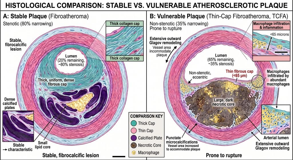
Histological comparison illustration. On the left, there is a stable fibroatheroma with an 80% narrowed lumen, a thick collagen fibrous cap, a small lipid core, and dense calcified plates. On the right, there is a vulnerable thin-cap fibroatheroma with a 35% non-stenotic lumen, extensive outward Glagov remodeling, a thin fibrous cap under 65 microns infiltrated by macrophages, and a large, dark necrotic core. The image was created with the help of artificial intelligence.
Plaque Rupture vs Plaque Erosion: Alternative Pathways to Thrombosis
Acute luminal thrombosis occurs through two distinct histopathological mechanisms:
| Feature | Plaque Rupture (~65–75% of Acute Coronary Events) | Plaque Erosion (~25–35% of Acute Coronary Events) |
| Fibrous Cap Integrity | Disrupted, torn, with a visible breach connecting the lumen to the necrotic core | Intact; no structural breach of the fibrous cap |
| Underlying Substrate | Large necrotic core, thin fibrous cap (< 65 µm), intense macrophage infiltration | Thick cap or absent necrotic core; base rich in extracellular matrix, versican, and hyaluronan |
| Trigger Mechanism | MMP-mediated collagen degradation; mechanical stress at plaque shoulder zones | Endothelial cell apoptosis and desquamation; high shear stress stripping the endothelial layer |
| Immune Drivers | Monocytes, macrophages, Th1 cells, NLRP3 inflammasome | Neutrophils, myeloperoxidase (MPO), Neutrophil Extracellular Traps (NETosis) |
| Patient Demographics | Older patients, males, strong association with hyperlipidemia and systemic inflammation | Younger patients, premenopausal females, smokers, absence of traditional multi-vessel CAD |
| Thrombus Composition | Fibrin-rich and erythrocyte-rich (“red thrombus”) | Platelet-rich (“white thrombus”) |
4. Atherosclerosis (Arterial Plaque) – Causes of Arterial Plaque Formation
Section Summary:
Arterial plaque forms from an obligate intersection between circulating atherogenic lipoproteins, localized endothelial barrier disruption, and an unresolved chronic inflammatory response. Without ApoB-containing particles penetrating the intimal space, atherosclerosis cannot develop regardless of arterial injury. Conversely, sustained particle retention initiates a self-reinforcing loop of lipid oxidation and immune activation that prevents tissue healing and drives chronic vascular architecture destruction.
Atherosclerosis (arterial plaque) is an etiologically complex disease. It does not arise from a single environmental pathogen or gene mutation, but rather from the intersection of three required biological conditions:
- The Presence of ApoB-Containing Lipoproteins: Atherosclerosis cannot develop in the absolute absence of ApoB lipoproteins. Regardless of systemic inflammatory status or vascular endothelial injury, an atheroma cannot form without ApoB particles crossing the endothelium and depositing sterols into the intimal matrix.
- Endothelial Permeability and Loss of Glycocalyx Integrity: The intact, healthy endothelium is an impermeable barrier to large macromolecular complexes. Chemical or physical disruptions—including low fluid shear stress, oxidative stress from tobacco toxins, and advanced glycation end-products—are required to allow lipoproteins to cross into the subendothelial space.
- Chronic, Unresolved Intimal Inflammation: Atheroma formation is not an acute reaction that heals with scarring. It is driven by failed pro-resolving lipid mediator signaling (deficits in resolvins, protectins, and maresins) combined with defective efferocytosis, locking the intima into a self-amplifying cycle of immune cell recruitment and tissue necrosis.
5. Atherosclerosis (Arterial Plaque) – Risk Factors for Arterial Plaque Formation
Section Summary:
Risk factors for atherosclerosis (arterial plaque) function as precise molecular drivers that accelerate intimal lipid accumulation, endothelial breakdown, or vascular inflammation. ApoB particle concentration reflects true atherogenic potential far more accurately than LDL cholesterol mass alone. Key contributors include genetically elevated Lipoprotein(a), hypertensive hydrostatic barotrauma, advanced glycation end-products in diabetes, particulate toxins from smoking, uremic milieu in renal failure, and somatic clonal hematopoiesis.
Risk factors for atherosclerosis (arterial plaque) do not operate through abstract epidemiological correlations; each driver accelerates specific biochemical steps within the vascular wall.
ApoB Particle Count vs LDL Cholesterol Mass
Traditional clinical medicine assesses cardiovascular risk using Low-Density Lipoprotein Cholesterol (LDL-C), which measures the total mass of cholesterol contained inside LDL particles (mg/dL or mmol/L).
However, pathophysiology confirms that the true driver of intimal particle deposition is Apolipoprotein B (ApoB)—or the absolute LDL Particle Number (LDL-P):
- Every atherogenic particle (LDL, VLDL, IDL, Lpa) carries exactly one molecule of ApoB-100.
- Plaque formation is governed by mass action: the probability that a particle enters and gets trapped within the subendothelial space is determined by the number of particles flowing through the lumen, not the cargo inside them.
- In conditions of metabolic dysfunction, insulin resistance, or hypertriglyceridemia, LDL particles become depleted of cholesteryl esters and enriched with triglycerides, generating small, dense LDL (sdLDL).
- Under these circumstances, a patient may have an apparently normal LDL-C of 90 mg/dL, yet harbor an elevated LDL-P and ApoB level. Because each small particle carries less cholesterol mass, many more individual particles are required to yield that 90 mg/dL total.
- These smaller particles more readily cross the endothelial barrier and bind with higher affinity to intimal proteoglycans.
Lipoprotein(a) [Lp(a)]: The Genetically Driven Risk Multiplier
Lp(a) is an LDL-like particle in which ApoB-100 is covalently linked via a disulfide bond to a specialized, polymorphic glycoprotein called apolipoprotein(a) [apo(a)]:
- Apo(a) shares structural homology with plasminogen, containing multiple looped peptide domains called kringle domains (specifically kringle IV and kringle V).
- Due to this structural mimicry, Lp(a) competitively binds to fibrin, inhibiting plasminogen activation and suppressing physiological fibrinolysis. This creates an inherently pro-thrombotic profile.
- In addition, Lp(a) serves as the primary plasma carrier for oxidized phospholipids (OxPL). When Lp(a) enters the intima, its OxPL cargo directly binds to endothelial and macrophage scavenger receptors, potently driving vascular inflammation and accelerating fibrocalcific aortic valve disease.
- Plasma concentrations of Lp(a) are over 85% to 90% genetically determined by the LPA gene locus, remaining largely unaffected by diet, exercise, and standard statin therapy.
Hypertension: Hemodynamic Barotrauma
Chronic elevations in systolic and pulse pressure accelerate atherogenesis through direct mechanical and hormonal pathways:
- Elevated hydrostatic pressure damages endothelial cell junctions, increasing permeability to circulating macromolecules.
- Pulsatile strain stimulates vascular endothelial and smooth muscle cells to overproduce Angiotensin II locally.
- Local Angiotensin II binds to the AT1 receptor, upregulating NADPH oxidase, which generates large quantities of superoxide anions.
- Superoxide reacts with endothelium-derived nitric oxide to form peroxynitrite, a cytotoxic reactive nitrogen species. This uncouples eNOS, abolishes protective vasodilation, and oxidizes local structural proteins.
For a deeper understanding of high blood pressure, please read our Hypertension article.
Type 2 Diabetes Mellitus and Hyperglycemia
Chronic metabolic dysfunction accelerates atheroma development via non-enzymatic glycation:
- Persistent elevations in intracellular glucose cause non-enzymatic condensation of glucose with vascular amino groups, forming Advanced Glycation End-products (AGEs) on long-lived structural proteins like collagen and elastin.
- AGEs irreversibly cross-link matrix collagen fibers, causing arterial stiffening and trapping lipoproteins within the wall.
- Circulating AGEs bind to the Receptor for Advanced Glycation End-products (RAGE) on endothelial cells and macrophages, activating NF-κB and driving sustained transcription of adhesion molecules, cytokines, and pro-thrombotic factors.
- Concurrently, hyperinsulinemia stimulates VSMC (vascular smooth muscle cell) migration and proliferation, while hyperglycemia blunts the body’s natural fibrinolytic systems by elevating Plasminogen Activator Inhibitor-1 (PAI-1).
For a deeper understanding of the pathophysiology of hyperglycemia, please read our Type 2 Diabetes Mellitus article.
Cigarette Smoking and Inhaled Pollutants
Inhaled combustion products, particularly acrolein, polycyclic aromatic hydrocarbons, and fine particulate matter (PM2.5), enter the pulmonary circulation and trigger systemic oxidative stress:
- These toxins oxidatively inactivate dimethylarginine dimethylaminohydrolase (DDAH), the enzyme responsible for degrading asymmetric dimethylarginine (ADMA).
- Accumulation of ADMA, an endogenous eNOS inhibitor, uncouples eNOS and eliminates baseline nitric oxide production.
- Smoking increases circulating levels of oxidized LDL, directly triggers platelet hyperreactivity, and shifts the endothelial hemostatic balance toward thrombosis by downregulating thrombomodulin and upregulating tissue factor.
Chronic Kidney Disease (CKD)
The uremic environment accelerates atherosclerosis through systemic biochemical derangements:
- Retention of protein-bound uremic toxins (e.g., indoxyl sulfate, p-cresyl sulfate) damages endothelial glycocalyx architecture and downregulates Klotho expression.
- Disrupted mineral metabolism (hyperphosphatemia and elevated Fibroblast Growth Factor 23 [FGF23]) accelerates osteogenic transdifferentiation of VSMCs (Vascular smooth muscle cells) via Runx2 upregulation, inducing rapid medial and intimal calcification.
- Chronic low-grade systemic inflammation and impaired reverse cholesterol transport accelerate foam cell transformation.
Please read our article about Chronic Kidney Disease for an extensive analysis of this medical condition.
Clonal Hematopoiesis of Indeterminate Potential (CHIP)
One of the most significant discoveries in modern cardiovascular science is CHIP—a condition where somatic mutations in hematopoietic stem cells confer a competitive growth advantage, leading to an expanded clonal population of leukocytes without overt hematologic malignancy:
- CHIP is primarily driven by age-related mutations in epigenetic regulator genes, including TET2, DNMT3A, and ASXL1.
- Individuals with CHIP have a two- to four-fold increase in the risk of coronary heart disease, independent of traditional lipid levels.
- Macrophages derived from TET2-mutated monocytes exhibit hyper-reactive NLRP3 inflammasome activation, producing elevated levels of IL-1β and IL-6 within the arterial plaque microenvironment, rapidly accelerating lesion destabilization.
6. Atherosclerosis (Arterial Plaque) – Downstream Clinical Manifestations and Disease Connections
Section Summary:
Atherosclerosis (arterial plaque) is a systemic vascular disease whose clinical manifestations depend on the specific arterial bed compromised by luminal stenosis or thrombotic occlusion. It directly causes coronary artery disease and heart failure through myocardial ischemia and tissue loss. Furthermore, it drives chronic kidney disease via microvascular damage and renovascular hypoperfusion, while producing disabling strokes, peripheral gangrene, and life-threatening abdominal aortic aneurysms.

Atherosclerosis (arterial plaque) is fundamentally a generalized systemic disease; an individual harboring advanced plaque in one arterial territory almost universally has subclinical or clinical disease in others. The specific clinical presentation is dictated by the vascular territory involved:
| Vascular Bed | Primary Target Arteries | Typical Clinical Manifestations | Primary Pathological Mechanism |
| Coronary Circulation | Left anterior descending, circumflex, right coronary artery | Stable angina, Unstable angina, NSTEMI, STEMI, Ischemic cardiomyopathy | Progressive luminal stenosis or acute plaque rupture/erosion with occlusive thrombus |
| Cerebrovascular Circulation | Internal carotid bulb, middle cerebral, basilar, vertebral | Transient ischemic attack (TIA), Non-cardioembolic ischemic stroke, Vascular dementia | Carotid bifurcation plaque rupture with distal thromboembolism or in situ intracranial thrombosis |
| Renal Circulation | Main renal arteries, intrarenal interlobar and arcuate arteries | Renovascular hypertension, Ischemic nephropathy, Accelerated CKD | Ostial renal artery stenosis causing renal hypoperfusion and hyperactivation of the RAAS cascade |
| Peripheral Circulation | Iliofemoral, superficial femoral, popliteal, tibial arteries | Intermittent claudication, Critical limb-threatening ischemia (rest pain, gangrene) | Fixed arterial stenosis restricting exercise hyperemia or acute thromboembolism |
| Aortic Circulation | Infrarenal abdominal aorta, thoracic aorta | Abdominal Aortic Aneurysm (AAA), Aortic atheroembolism (blue toe syndrome) | Intimal inflammation driving medial elastolysis and wall thinning; mechanical dilation |
Detailed Disease Connections
Coronary Artery Disease (CAD)
Coronary Artery Disease is the direct clinical consequence of coronary atherosclerosis (arterial plaque). It presents across two clinical spectra:
- Chronic Coronary Syndromes (Stable CAD): Characterized by high-grade, structurally stable fibroatheromas that narrow the coronary lumen by > 70%. Under conditions of increased myocardial oxygen demand (e.g., exercise or emotional stress), coronary flow reserve is exhausted, producing transient subendocardial ischemia perceived clinically as stable angina pectoris.
- Acute Coronary Syndromes (ACS): Characterized by the acute disruption of a vulnerable plaque (either rupture of a thin fibrous cap or endothelial erosion). Exposure of the thrombogenic necrotic core triggers platelet aggregation and fibrin deposition, forming an intraluminal thrombus. Incomplete or transient occlusion produces Unstable Angina or Non-ST-Segment Elevation Myocardial Infarction (NSTEMI). Complete, persistent occlusion produces ST-Segment Elevation Myocardial Infarction (STEMI), leading to transmural myocardial necrosis within hours if it is left untreated.
Congestive Heart Failure (CHF)
Atherosclerosis (arterial plaque) is the single most common etiology of Congestive Heart Failure (CHF), operating through two distinct pathways:
- Heart Failure with Reduced Ejection Fraction (HFrEF / Ischemic Cardiomyopathy): Myocardial infarctions cause irreversible necrosis of contractile cardiomyocytes. Because adult cardiomyocytes have minimal regenerative capacity, necrotic myocardium is replaced by non-contractile fibrillar collagen scars. The remaining viable myocardium undergoes maladaptive hypertrophy and spherical dilation (ventricular remodeling), reducing left ventricular systolic ejection fraction (< 40%).
- Heart Failure with Preserved Ejection Fraction (HFpEF): Chronic systemic atherosclerosis and its associated risk factors (hypertension, diabetes, obesity) produce systemic microvascular endothelial inflammation. Endothelial cells in coronary microvessels express low levels of eNOS, which reduces protein kinase G (PKG) activity in adjacent cardiomyocytes. This leads to cardiomyocyte stiffening and interstitial fibrosis, impairing diastolic relaxation and elevating left ventricular filling pressures despite a preserved ejection fraction (> 50%).
Chronic Kidney Disease (CKD)
Atherosclerosis (arterial plaque) and Chronic Kidney Disease (CKD) are linked through a bidirectional cardiorenal axis:
- Atherosclerotic Renal Artery Stenosis (ARAS): Plaque accumulation at the ostium of the main renal artery restricts renal blood flow, which decreases perfusion pressure at the afferent arteriole of the juxtaglomerular apparatus. This triggers constant secretion of renin, activating the Renin-Angiotensin-Aldosterone System (RAAS). Angiotensin II induces systemic vasoconstriction, while aldosterone promotes renal sodium and water retention, producing renovascular hypertension and chronic ischemic atrophy of renal tubules.
- Intrarenal Arteriosclerosis and Glomerulosclerosis: Microvascular atheroma and hyaline arteriolosclerosis within the interlobular arteries and afferent arterioles cause chronic glomerular ischemia. Glomeruli undergo global collapse, sclerosis, and nephron loss, accelerating the loss of estimated Glomerular Filtration Rate (eGFR).
- The Uremic Acceleration Loop: Declining kidney function impairs the clearance of phosphate and uremic toxins. This directly triggers osteochondrogenic phenotypic transdifferentiation of vascular smooth muscle cells, creating extensive vascular calcification that worsens systemic atherosclerosis.
7. Prevention of Atherosclerosis (Arterial Plaque)
Section Summary:
Prevention of atherosclerosis (arterial plaque) requires a tiered clinical strategy addressing lifetime exposure to atherogenic lipoproteins and vascular stressors. Primordial prevention preserves optimal endothelial function from youth, while primary prevention targets asymptomatic plaque progression using lifestyle interventions and early risk stratification. Secondary prevention stabilizes established plaques to avoid recurrent thrombotic events through intensive lipid reduction, glycemic optimization, blood pressure normalization, and smoking cessation.

Cardiovascular prevention is categorized across three clinical stages:
- Primordial Prevention: Preventing the initial acquisition of cardiovascular risk factors (e.g., maintaining lifetime normal blood pressure, optimal body composition, and low ApoB levels from childhood).
- Primary Prevention: Treating identified risk factors in individuals who have not yet developed clinical cardiovascular events or flow-limiting disease, preventing the progression of subclinical plaque into clinical crises.
- Secondary Prevention: Aggressively treating individuals with established clinical atherosclerotic disease (prior MI, stroke, revascularization) to prevent recurrent ischemic events, plaque rupture, and death.
The Lifetime Cumulative Exposure Paradigm: The “Area Under the Curve”
Atherosclerosis (arterial plaque) does not develop from short-term exposure to risk factors. It is governed by cumulative lifetime exposure, often conceptualized as the “area under the curve” for atherogenic particles (e.g., circulating ApoB or LDL-C concentration multiplied by age, expressed in gram-years):
- A person with a moderate LDL-C of 130 mg/dL maintained over 50 years accumulates an equivalent biological intimal lipid exposure as an individual with Familial Hypercholesterolemia harboring an LDL-C of 260 mg/dL over 25 years.
- Consequently, lowering atherogenic particles early in life provides greater relative risk reduction than starting identical therapy late in life after extensive necrotic cores have formed.
Lifestyle and Dietary Interventions
Lifestyle modifications form the cornerstone of all prevention stages:
- Dietary Patterns: Diets rich in minimally processed whole foods, high in dietary soluble fiber, and low in ultra-processed carbohydrates and saturated fats significantly reduce cardiovascular risk. Soluble viscous fibers (e.g., beta-glucan, psyllium) bind bile acids in the intestinal lumen, increasing fecal bile acid excretion. This depletes hepatic cholesterol stores and upregulates hepatic LDL receptors, lowering circulating ApoB particles.
- Replacement of Saturated Fatty Acids: Replacing saturated fatty acids (which downregulate hepatic LDLR by altering hepatic membrane fluidity) with polyunsaturated (PUFA) and monounsaturated (MUFA) fatty acids restores hepatic LDLR expression and lowers circulating atherogenic particles.
- Exercise Physiology: Regular aerobic and resistance exercise increases laminar blood flow and sustained pulsatile shear stress throughout the arterial tree. This activates endothelial mechanosensors, upregulates KLF2, increases eNOS expression, and elevates baseline nitric oxide bioavailability. Exercise also improves peripheral insulin sensitivity, lowers resting sympathetic tone, and reduces systemic inflammatory cytokines.
- Complete Tobacco and Nicotine Cessation: Smoking cessation rapidly reverses acute endothelial dysfunction and platelet hyperreactivity within days to weeks. Over years, it halts the accelerated accumulation of intimal lipids, reducing cardiovascular event rates by 50% within one to two years after cessation.
8. Treatment of Atherosclerosis (Arterial Plaque): Medical Therapy, Plaque Stabilization, and Revascularization
Section Summary:
Modern treatment of atherosclerosis focuses on aggressive lipid lowering, inflammation suppression, and plaque stabilization rather than mechanical eradication. Pharmacotherapies such as statins, ezetimibe, and PCSK9 inhibitors deplete lipid cores and thicken fibrous caps to prevent rupture. In advanced flow-limiting disease, revascularization via percutaneous coronary intervention or bypass surgery restores downstream perfusion without eliminating the underlying systemic disease.

A common patient misconception is that medical treatment “melts away” or “vacuums” plaque out of the arterial wall. Clinical imaging trials utilizing high-resolution Intravascular Ultrasound (IVUS) and Optical Coherence Tomography (OCT) demonstrate that the primary goal of medical therapy is plaque stabilization, accompanied by modest plaque regression.
Plaque Stabilization vs Plaque Regression
Aggressive medical therapy transforms a lethal, rupture-prone plaque into a safe, quiescent lesion through specific structural changes:
- Lipid Core Depletion: Lowering circulating ApoB lipoproteins creates a concentration gradient that draws unesterified cholesterol out of the intimal space, shrinking the volume of the necrotic lipid core.
- Fibrous Cap Thickening: Suppression of intimal inflammation reduces macrophage secretion of MMPs (matrix metalloproteinases). Living synthetic VSMCs (Vascular Smooth Muscle Cells) continue to synthesize collagen, increasing the thickness of the fibrous cap well beyond the critical 65 µm rupture threshold.
- Macrocalcific Consolidation: Under statin therapy, spotty microcalcifications coalesce into dense, continuous macrocalcific plates. This stabilizes the lesion mechanically and protects against fibrous cap rupture.
| Drug Class | Representative Agents | Primary Molecular Mechanism | Effect on Plaque Biology | Clinical Endpoints Demonstrated |
| Statins (HMG-CoA Reductase Inhibitors) | Atorvastatin, Rosuvastatin | Competitively inhibit HMG-CoA reductase; deplete hepatic cholesterol; upregulate hepatic LDL receptors | Decreases necrotic core volume; thickens fibrous cap; promotes macrocalcification; reduces hs-CRP | Substantial reduction in MI, stroke, cardiovascular mortality (4S, WOSCOPS, JUPITER) |
| Cholesterol Absorption Inhibitors | Ezetimibe | Inhibits Niemann-Pick C1-Like 1 (NPC1L1) transporter at the enterocyte brush border | Reduces intimal ApoB influx; provides additive lipid lowering when combined with statins | Further reduces cardiovascular events when added to statin therapy (IMPROVE-IT) |
| PCSK9 Monoclonal Antibodies | Evolocumab, Alirocumab | Binds circulating PCSK9 protein, preventing PCSK9-mediated degradation of hepatic LDL receptors | Induces profound LDL-C lowering (50–60%); leads to measurable atheroma regression on IVUS | Significant reduction in MI, stroke, and revascularization in high-risk patients (FOURIER, ODYSSEY) |
| PCSK9 Small Interfering RNA (siRNA) | Inclisiran | Cleaves PCSK9 mRNA inside hepatocytes via the RNA-induced silencing complex (RISC) | Provides sustained inhibition of PCSK9 synthesis with twice-yearly dosing; lowers circulating ApoB | Demonstrated sustained 50% LDL-C reduction (ORION clinical trial program) |
| Adenosine Triphosphate-Citrate Lyase (ACL) Inhibitors | Bempedoic Acid | Inhibits ACL upstream of HMG-CoA reductase in the hepatic cholesterol synthesis pathway | Depletes hepatic cholesterol and upregulates LDL receptors without causing muscular side effects | Lowers LDL-C and reduces major adverse cardiovascular events in statin-intolerant patients (CLEAR Outcomes) |
| Purified Omega-3 Fatty Acids | Icosapent Ethyl | High-dose pure ethyl ester of eicosapentaenoic acid (EPA); incorporates into cell membranes | Stabilizes endothelial membranes; inhibits lipid oxidation; reduces macrophage activation | Significant 25% relative risk reduction in major adverse cardiovascular events (REDUCE-IT) |
| Anti-Inflammatory Therapies | Colchicine (low-dose, 0.5 mg daily) | Inhibits tubulin polymerization; disrupts microtubule assembly; suppresses NLRP3 inflammasome | Prevents cholesterol crystal-induced release of IL-1β and IL-18; halts cap-cleaving inflammation | Significant reduction in recurrent ischemic cardiovascular events (COLCOT, LoDoCo2) |
| Antiplatelet Agents | Aspirin, Clopidogrel, Ticagrelor | Aspirin irreversibly inhibits COX-1 (blocking Thromboxane A2); P2Y12 inhibitors block ADP receptors | Prevents platelet aggregation and acute luminal thrombus formation if a plaque ruptures | Essential for preventing acute arterial occlusion in secondary prevention |
SGLT2 Inhibitors and GLP-1 Receptor Agonists
Originally developed for glycemic control in Type 2 Diabetes, Sodium-Glucose Cotransporter 2 (SGLT2) inhibitors (e.g., Empagliflozin, Dapagliflozin) and Glucagon-Like Peptide-1 (GLP-1) receptor agonists (e.g., Semaglutide, Dulaglutide) have transformed cardiovascular therapeutics:
- SGLT2 inhibitors reduce intraglomerular pressure, promote osmotic diuresis, lower systemic vascular resistance, and shift myocardial metabolism toward ketone bodies, demonstrating reductions in heart failure hospitalizations and Chronic Kidney Disease progression (EMPA-REG, DAPA-HF).
- GLP-1 receptor agonists directly reduce vascular inflammation, improve endothelial nitric oxide bioavailability, decrease systemic PAI-1, and reduce body weight, significantly lowering rates of non-fatal myocardial infarction and stroke (LEADER, SUSTAIN-6, SELECT).
Revascularization Strategies: Indications and Limitations
When atherosclerosis causes severe flow-limiting luminal stenosis or triggers acute thrombosis, revascularization restores blood supply:
- Percutaneous Coronary Intervention (PCI): Involves advancing a catheter to the coronary stenosis, dilating the lesion with a balloon (angioplasty), and deploying a drug-eluting stent (DES). Modern stents elute antiproliferative agents (e.g., everolimus, zotarolimus) to prevent smooth muscle hyperplasia and in-stent restenosis. In acute myocardial infarction (STEMI), emergency PCI of the culprit lesion is life-saving. However, in stable Coronary Artery Disease, multiple randomized trials (COURAGE, ISCHEMIA) demonstrate that PCI relieves angina but does not reduce the incidence of myocardial infarction or death compared to optimal medical therapy alone. This is because PCI addresses only the local flow-limiting lesion, leaving systemic, non-obstructive vulnerable plaques elsewhere in the arterial tree untreated.
- Coronary Artery Bypass Grafting (CABG): A surgical procedure in which conduits (internal mammary arteries, saphenous veins) are grafted to bypass diseased arterial segments. CABG provides superior survival outcomes compared to PCI in patients with complex multi-vessel disease, left main coronary disease, or diabetic patients with multi-vessel Coronary Artery Disease. CABG acts as a biological buffer: by delivering blood flow distal to the mid-vessel segments, it protects downstream myocardium not only from current stenosis but also from future plaque ruptures occurring proximal to the graft anastomosis.
9. High-Yield Frequently Asked Questions (FAQs) about Atherosclerosis (Arterial Plaque)
Section Summary:
Frequently asked clinical questions address practical patient concerns regarding plaque reversibility, imaging interpretations, and risk misconceptions. Key topics include understanding why normal stress test results do not rule out impending myocardial infarction, how a coronary artery calcium score guides preventive decisions, why statins increase plaque calcification as a healing response, and the systemic nature of vascular disease across distinct organs.
Can arterial plaque ever completely disappear?
Arterial plaque does not completely disappear once established. An advanced atheroma is a complex structural alteration consisting of a necrotic core, cell fragments, collagenous scar tissue, and mineral calcification.
Intravascular imaging studies (such as the ASTEROID and GLAGOV trials) demonstrate that intensive medical therapy can produce modest atheroma volume regression (typically shrinking total plaque volume by 1% to 3% per year). However, the primary clinical goal of treatment is plaque stabilization: shrinking the lipid core, thickening the protective fibrous cap, and halting local inflammation so the plaque becomes an inert, rupture-resistant scar.
Why was my stress test normal if I had a heart attack two weeks later?\
An exercise stress test evaluates whether an arterial blockage restricts blood flow during physical exertion (an oxygen supply-demand mismatch). A stress test typically becomes abnormal only when an arterial stenosis occludes 70% or more of the vessel lumen.
As demonstrated by the Glagov phenomenon, most acute myocardial infarctions are caused by the sudden rupture of non-obstructive, vulnerable plaques that narrow the lumen by only 30% to 50%. Because these mild blockages do not restrict exercise-induced blood flow, the stress test is completely normal. However, because these lesions harbor thin fibrous caps and large, inflamed necrotic cores, they can tear open unpredictably, generating an acute occlusive thrombus.
My doctor said my coronary calcium score went up after starting a statin. Did the medication worsen my plaque?
No. An increase in the Coronary Artery Calcium (CAC) score after initiating statin therapy is an expected biological response reflecting plaque stabilization, not disease progression.
Statins deplete the inflammatory, pro-thrombotic lipid core and stimulate vascular smooth muscle cells to lay down dense macrocalcifications. This process transforms fragile, dangerous microcalcifications into solid calcific plates that physically lock the plaque in place and prevent rupture. While your score rises because CT scans detect this denser mineral footprint, the underlying biological hazard of the plaque has significantly decreased.
Is an LDL cholesterol test alone enough to assess my plaque risk?
No. Standard lipid panels measure Low-Density Lipoprotein Cholesterol (LDL-C), which reflects the total mass of cholesterol within LDL particles. Under conditions of metabolic dysfunction, prediabetes, or elevated triglycerides, the body produces high quantities of small, dense LDL particles (sdLDL).
Each small particle carries less cholesterol mass, meaning a patient can have an apparently normal LDL-C concentration despite harboring an elevated LDL Particle Number (LDL-P) and Apolipoprotein B (ApoB) concentration. Because plaque formation is driven by the absolute number of particles penetrating and becoming trapped within the intimal wall, measuring ApoB or LDL-P provides a far more accurate assessment of atherogenic risk than LDL-C alone.
If I have plaque in my legs (PAD), does that mean I have plaque in my heart?
Yes. Atherosclerosis is a systemic, generalized vascular pathology. If an individual has accumulated enough plaque in the peripheral arterial tree to cause symptoms like intermittent claudication or abnormal ankle-brachial indices, extensive subclinical or clinical atherosclerosis is almost certainly present in the coronary and carotid circulations. Patients diagnosed with PAD face cardiovascular mortality rates comparable to those of patients who have already suffered a myocardial infarction.
Can “chelation therapy” or over-the-counter supplements clean out my arteries?
No. There is no rigorous scientific evidence that over-the-counter supplements, herbal cleanses, or oral chelation agents dissolve or eliminate arterial plaque. Plaque resides inside the arterial wall beneath the endothelial cell monolayer; it is not exposed to the luminal stream and cannot be washed away.
Treatments marketed directly to consumers claiming to clean or flush out arteries are scientifically unfounded and dangerous when they lead patients to delay or discontinue evidence-based medical therapies.
What is the difference between a conventional coronary angiogram and a coronary CT angiogram (CCTA)?
A conventional coronary angiogram is an invasive, catheter-directed fluoroscopic luminogram. It visualizes the silhouette of contrast within the arterial lumen, identifying areas of severe, flow-limiting stenosis, but it cannot visualize the arterial wall itself or non-obstructive, positively remodeled plaques.
A Coronary Computed Tomography Angiogram (CCTA) is a non-invasive scan that uses intravenous contrast and advanced multi-detector CT imaging to visualize both the lumen and the arterial wall. CCTA can identify non-obstructive plaques, evaluate the extent of positive outward Glagov remodeling, quantify the necrotic core, and identify high-risk plaque features like low-attenuation plaque, napkin-ring signs, and spotty calcification long before a lesion causes luminal stenosis.
10. High-Yield Questions about Atherosclerosis (Arterial Plaque) to Ask Your Doctor
Section Summary:
Effective communication between patients and clinicians requires targeted, evidence-based inquiries regarding individual vascular vulnerability and therapeutic goals. Patients should ask specific questions about their cumulative ApoB or LDL particle count, coronary calcium assessment, plaque morphology on advanced imaging, individualized lipid-lowering targets, and personalized anti-inflammatory strategies to ensure comprehensive protection beyond traditional broad-spectrum risk score estimations.

When discussing cardiovascular risk and plaque management with your physician, the following targeted clinical questions can guide a precise, personalized prevention and treatment plan:
- What is my absolute Apolipoprotein B (ApoB) particle count? Ask whether your ApoB level is concordant with your standard LDL-C, particularly if you have elevated triglycerides, metabolic syndrome, or a family history of premature heart disease.
- Have I ever had my Lipoprotein(a) [Lp(a)] level measured? Because Lp(a) is an independent, genetically determined risk multiplier that is not captured on standard cholesterol panels, request a one-time, lifetime screening measurement.
- Would I benefit from a non-contrast Coronary Artery Calcium (CAC) scan or a Coronary CT Angiogram (CCTA)? If your cardiovascular risk category is intermediate or uncertain, ask whether anatomical imaging can directly quantify your plaque burden and reclassify your treatment intensity.
- What is my individualized LDL-C or ApoB target? Guidelines recommend distinct lipid-lowering targets based on absolute risk (e.g., an LDL-C target < 55 mg/dL (< 1.4 mmol/L) or ApoB < 65 mg/dL for individuals with established vascular disease or high risk, versus higher thresholds for low-risk primary prevention). Ask what your specific numerical target is.
- What is my systemic inflammatory status as measured by high-sensitivity C-reactive protein (hs-CRP)? Inquiring about your hs-CRP helps determine whether you have residual inflammatory risk that warrants targeted anti-inflammatory therapy (e.g., low-dose colchicine).
- If an arterial blockage is found, does it require a stent, or should we optimize my medical therapy first? If you have stable symptoms without acute coronary syndrome, ask about the evidence from trials like ISCHEMIA, which demonstrate that aggressive medical therapy matches stent placement in preventing death and future myocardial infarctions.
- Are there specific non-statin therapies that should be combined with my current regimen? If your lipid targets are not reached with maximally tolerated statin therapy, ask whether adding an agent with a complementary mechanism (such as ezetimibe or a PCSK9 inhibitor) is indicated.
11. Credible, Peer-Reviewed, High-Trust Sources
The scientific principles detailed throughout this clinical reference are founded on the following landmark clinical trials, pathological investigations, and international consensus guidelines:
Glagov, S., et al. (1987). “Compensatory enlargement of human atherosclerotic coronary arteries.” The New England Journal of Medicine, 316(22), 1371–1375.
- The landmark human autopsy study demonstrating outward arterial remodeling (the Glagov phenomenon), proving that arteries enlarge to prevent luminal narrowing until plaque occupies more than 40% of the internal elastic lamina area.
Tabas, I., & Bornfeldt, K. E. (2016). “Macrophage Phenotype and Function in Different Stages of Atherosclerosis.” Circulation Research, 118(4), 653–667.
- Authoritative pathological review detailing the molecular mechanisms of foam cell formation, defective efferocytosis via MerTK downregulation, and secondary necrosis in the formation of the necrotic core.
Ridker, P. M., et al. (2017). “Antiinflammatory Therapy with Canakinumab for Atherosclerotic Disease.” The New England Journal of Medicine, 377(12), 1119–1131. (The CANTOS Trial)
- Landmark randomized, double-blind trial demonstrating that selectively inhibiting the IL-1β to IL-6 inflammatory pathway significantly reduces recurrent cardiovascular event rates without altering circulating lipid levels, validating the inflammatory hypothesis of atherosclerosis.
Sabatine, M. S., et al. (2017). “Evolocumab and Clinical Outcomes in Patients with Cardiovascular Disease.” The New England Journal of Medicine, 376(18), 1713–1722. (The FOURIER Trial)
- Pivotal phase 3 trial establishing that intensive LDL-C lowering to median levels of 30 mg/dL using the PCSK9 inhibitor evolocumab significantly reduces major adverse cardiovascular events without plateauing benefit.
Nissen, S. E., et al. (2006). “Effect of very high-intensity statin therapy on regression of coronary atherosclerosis: the ASTEROID trial.” JAMA, 295(13), 1556–1565.
- Prospective intravascular ultrasound (IVUS) imaging trial demonstrating that intensive lipid-lowering therapy with rosuvastatin can induce measurable regression of coronary atheroma volume.
Tardif, J. C., et al. (2019). “Efficacy and Safety of Low-Dose Colchicine after Myocardial Infarction.” The New England Journal of Medicine, 381(26), 2497–2505. (The COLCOT Trial)
- Landmark outcome trial proving that broad-spectrum anti-inflammatory therapy with low-dose colchicine (0.5 mg daily) significantly lowers the risk of ischemic cardiovascular events in patients with recent myocardial infarction.
Mach, F., et al. (2019 / 2020). “2019 ESC/EAS Guidelines for the management of dyslipidaemias: lipid modification to reduce cardiovascular risk.” European Heart Journal, 41(1), 111–188.
- European clinical practice guidelines outlining risk-stratified treatment thresholds, emphasizing aggressive LDL-C targets (< 55 mg/dL for very-high-risk patients) and the clinical utility of ApoB and non-invasive imaging.
Grundy, S. M., et al. (2018 / 2019). “2018 AHA/ACC/AACVPR/AAPA/ABC/ACPM/ADA/AGS/APhA/ASPC/NLA/PCNA Guideline on the Management of Blood Cholesterol.” Circulation, 139(25), e1082–e1143.
- Comprehensive American multi-society consensus guidelines establishing recommendations for statin and non-statin therapies, CAC scoring for risk reclassification, and secondary prevention algorithms.
Featured Image: 3D biomedical cross-section of a muscular artery showing intramural atherosclerotic plaque with a lipid necrotic core, microcalcifications, and fibrous cap beneath the endothelium, created with the help of Artificial Intelligence.
© UncutMed — Medical knowledge, precise, truthful, unfiltered. The current article was last modified on October 7, 2026. This article was clinically authored and reviewed under international evidence-based standards. Always consult your personal healthcare physician before modifying any medical therapy.