© UncutMed — Medical knowledge, precise, truthful, unfiltered. The current article was last modified on September 16, 2026. This article was clinically authored and reviewed under international evidence-based standards. Always consult your personal healthcare physician before modifying any medical therapy.
Medical Disclaimer: This article is for informational and educational purposes only. It does not constitute medical advice, formal diagnosis, or treatment recommendations. Always consult a qualified healthcare professional regarding any metabolic condition or therapeutic plan.
1. Medical Definition of Chronic Kidney Disease
Section Summary:
Chronic Kidney Disease (CKD) is clinically defined as abnormalities of kidney structure or function persisting for more than three months, with health implications. Diagnostic criteria require either a decreased glomerular filtration rate (GFR < 60 mL/min/1.73 m²) or one or more verified markers of kidney damage, such as persistent albuminuria.
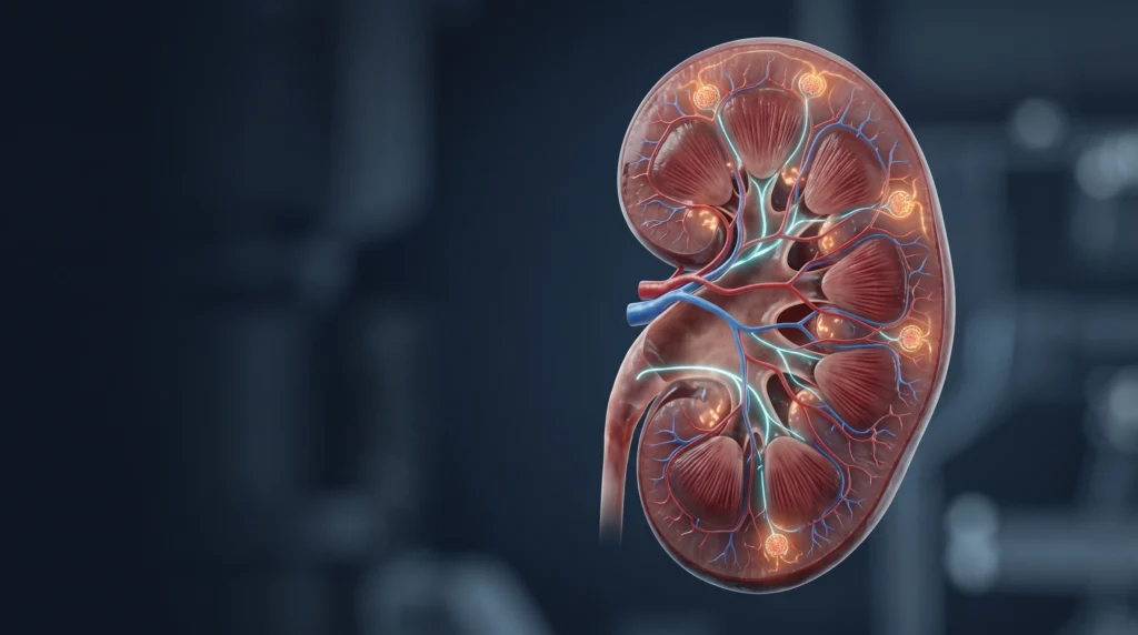
Chronic Kidney Disease (CKD) is not a single disease entity, but rather a pathophysiological syndrome characterized by progressive, irreversible loss of functional renal parenchyma. The global diagnostic standard, established by the Kidney Disease: Improving Global Outcomes (KDIGO) consensus, defines Chronic Kidney Disease as abnormalities of kidney structure or function persisting for more than 3 months, with implications for health.
The requirement for a minimum 3-month duration (“chronicity”) is vital to distinguish Chronic Kidney Disease from Acute Kidney Injury (AKI) or Acute Kidney Diseases and Disorders (AKD), which are potentially reversible.
Under KDIGO criteria, an individual is diagnosed with CKD if they demonstrate either of the following criteria present for more than 3 months:
- Decreased GFR: Glomerular filtration rate (GFR)< 60 mL/min/1.73 m². A GFR below this threshold represents a loss of more than half of the normal adult kidney filtration capacity, at which point the prevalence of metabolic, endocrine, and cardiovascular complications rises steeply.
- Markers of Kidney Damage (One or more):
- Albuminuria: Urine Albumin-to-Creatinine Ratio (uACR) ≥ 30 mg/g (≥ 3 mg/mmol).
- Urine sediment abnormalities: Persistent presence of dysmorphic red blood cells, acanthocytes, red blood cell casts, white blood cell casts, or broad waxy casts.
- Electrolyte and other abnormalities due to tubular disorders (e.g., renal tubular acidosis, nephrogenic diabetes insipidus).
- Histological abnormalities: Diagnostic changes demonstrated on percutaneous renal biopsy (e.g., glomerulosclerosis, tubulointerstitial fibrosis, immune complex deposition).
- Structural abnormalities: Detected via kidney ultrasonography, CT, or MRI (e.g., polycystic kidneys, renal cortical thinning, renal artery stenosis, hydronephrosis).
- History of Kidney Transplantation: Recipients are classified as having Chronic Kidney Disease regardless of their baseline allograft function, due to the underlying presence of structural and immunological damage.
2. Chronic Kidney Disease Classification and Staging
Section Summary:
Chronic Kidney Disease is staged using the international KDIGO “CGA” classification system based on Cause (C), GFR category (G1–G5), and Albuminuria category (A1–A3). This two-dimensional matrix categorizes patients into Low, Moderate, High, or Very High risk for renal disease progression, cardiovascular mortality, and all-cause mortality.
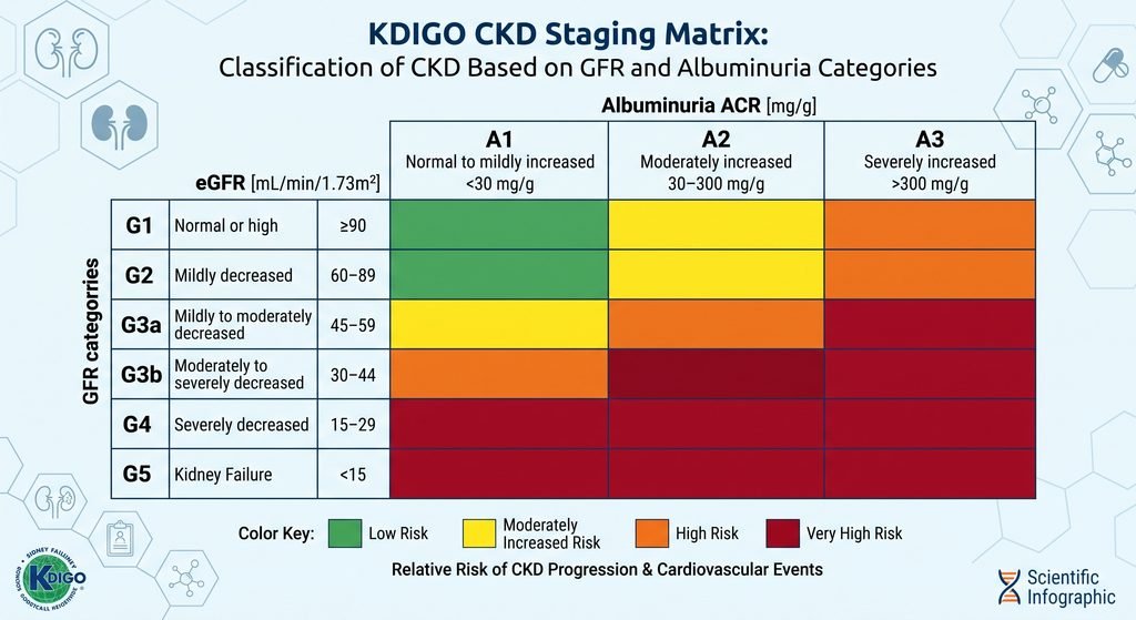
The KDIGO staging system uses a two-dimensional grid combining filtration function (GFR, stages G1 to G5) with vascular and podocyte barrier integrity (Albuminuria, stages A1 to A3). Identifying the underlying etiology (Cause) provides the full clinical profile (CGA classification).
GFR Staging
| GFR Stage | eGFR (mL/min/1.73 m²) | Clinical Description | Pathological State |
| G1 | ≥ 90 | Normal or high | Normal filtration; requires structural or urinary marker for CKD diagnosis |
| G2 | 60-89 | Mildly decreased | Mild functional loss relative to young adult normal; requires damage marker |
| G3a | 45-59 | Mildly to moderately decreased | True functional insufficiency; early hormonal/mineral imbalances emerge |
| G3b | 30-44 | Moderately to severely decreased | Advanced insufficiency; accelerated cardiorenal risk, anemia, bone disease |
| G4 | 15-29 | Severely decreased | Pre-end-stage renal failure; preparation for kidney replacement therapy |
| G5 | < 15 | Kidney failure (End-Stage Renal Disease / ESRD) | Overt uremia; requires dialysis or preemptive kidney transplantation |
Albuminuria Staging
| Albuminuria Stage | uACR (mg/g) | uACR (mg/mmol) | Clinical Description | Pathological Correlate |
| A1 | < 30 | < 3 | Normal to mildly increased | Intact podocyte slit diaphragms and filtration barrier |
| A2 | 30-300 | 3-30 | Moderately increased (formerly called microalbuminuria) | Subclinical endothelial dysfunction, early podocyte effacement |
| A3 | > 300 | > 30 | Severely increased (formerly called macroalbuminuria) | Extensive basement membrane disruption, nephrotic-range leakage |
The Prognostic Heatmap (CGA Risk Stratification)
The intersection of GFR and Albuminuria categories directly dictates clinical trajectory:
- Green (Low Risk): Stage G1/G2 with A1. This is not classified as Chronic Kidney Disease unless structural or genetic kidney lesions are verified.
- Yellow (Moderately Increased Risk): G3a with A1 or G1/G2 with A2. Warrants annual monitoring of eGFR and uACR.
- Orange (High Risk): G3b with A1, G3a with A2, or G1/G2 with A3. Requires evaluation every 6 months and active pharmacotherapy to prevent disease progression.
- Red (Very High Risk): Stage G4 or G5 across all albuminuria levels, or Stage G3b with A2–A3. Requires management by a nephrologist and multidisciplinary care.
3. Chronic Kidney Disease Pathophysiology (Extensive Analysis)
Section Summary:
Chronic Kidney Disease progression follows a common pathophysiological pathway independent of the initial insult. Nephron loss triggers compensatory glomerular hyperfiltration, hemodynamic shear stress, renin-angiotensin-aldosterone system overactivation, proteinuria-induced tubulointerstitial inflammation, and microvascular rarefaction, culminating in progressive tubulointerstitial fibrosis and glomerulosclerosis.
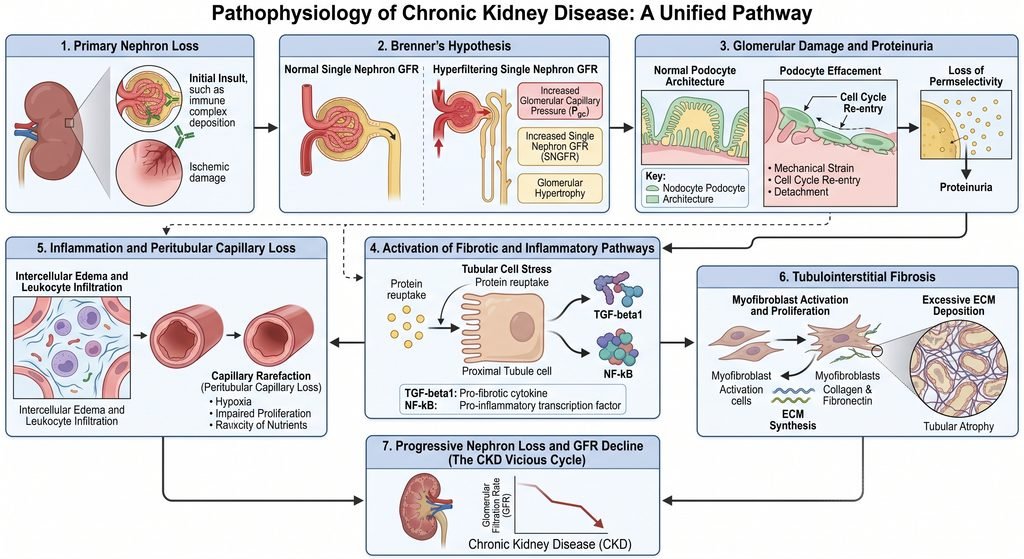
The pathophysiology of Chronic Kidney Disease follows these steps:
- Initial Insult (e.g., Diabetes, Hypertension, Glomerulonephritis)
- Primary Nephron Loss
- Compensatory Glomerular Hyperfiltration (Brenner Hypothesis: ↑ Angiotensin II → Efferent Constriction)
- Intraglomerular Hypertension & Shear Stress
- Podocyte Effacement, Detachment & Proteinuria Leakage
- Tubular Cell Uptake of Albumin & Toxic Solutes
- Release of Pro-inflammatory (NF-κB, MCP-1) & Pro-fibrotic (TGF-β1) Factors
- Peritubular Capillary Rarefaction & Tubulointerstitial Fibrosis
- Global Glomerulosclerosis & End-Stage Renal Disease (ESRD)
The pathophysiology of Chronic Kidney Disease involves both the initial disease process and self-perpetuating secondary injury mechanisms. When renal parenchymal injury destroys a critical mass of nephrons, the remaining viable nephrons undergo functional and structural changes to preserve total renal clearance. While acutely compensatory, these adaptations become maladaptive over time and drive progressive renal decline.
1. The Brenner Hypothesis: Compensatory Hyperfiltration and Glomerular Hemodynamics
Barry Brenner’s hyperfiltration hypothesis remains a foundational concept in renal pathophysiology:
- Nephron Loss and Structural Adaptation: As nephrons are destroyed, the total renal filtration surface area decreases. To maintain overall homeostatic filtration, the remaining intact nephrons undergo compensatory hypertrophy.
- Hemodynamic Alterations: Glomerular plasma flow and single-nephron GFR (snGFR) increase. This state is mediated by profound vasodilation of the afferent arteriole combined with angiotensin II-induced vasoconstriction of the efferent arteriole.
- Intraglomerular Capillary Hypertension: The imbalance between afferent entry and efferent exit pressures creates elevated intraglomerular hydraulic pressure. This increases transcapillary filtration of fluid, driving compensatory hyperfiltration.
- Biomechanical Shear Stress and Podocyte Injury: The elevated pressure exposes the delicate glomerular capillary tuft to chronic mechanical shear stress. Visceral epithelial cells (podocytes) are terminally differentiated and cannot divide to replace lost cells. Persistent stretch causes podocyte hypertrophy, foot process effacement, detachment from the glomerular basement membrane (GBM), and denudation of the capillary wall.
- Focal Segmental to Global Glomerulosclerosis: The exposed basement membrane adheres to Bowman’s capsule (synechia formation), initiating focal segmental glomerulosclerosis (FSGS). Plasma macromolecules leak into the subendothelial space (hyalinosis), which eventually consolidates into global glomerulosclerosis, destroying the nephron entirely.
2. Proteinuria as an Independent Cytotoxic and Fibrotic Driver
Filtered urinary proteins are directly involved in downstream nephrotoxicity, rather than simply being passive biomarkers of glomerular barrier breakdown:
- Tubular Albumin Overload: Under high intraglomerular pressure and podocyte loss, high volumes of plasma proteins (albumin, complement factors, transferrin, immunoglobulins) cross into the proximal convoluted tubule.
- Receptor-Mediated Endocytosis: The proximal tubule epithelium reabsorbs filtered proteins via the megalin-cubilin receptor complex. In proteinuric states, this transport system becomes overwhelmed.
- Intracellular Signaling and Inflammation: Intracellular protein overload induces endoplasmic reticulum (ER) stress, lysosomal rupture, and oxidative stress via reactive oxygen species (ROS) generation. This activates transcription factors such as Nuclear Factor-kappa B (NF-κB), triggering the production of pro-inflammatory chemokines, particularly Monocyte Chemoattractant Protein-1 (MCP-1), Interleukin-6 (IL-6), and Interleukin-8 (IL-8).
- Myofibroblast Transformation and Collagen Deposition: Macrophages and T-lymphocytes infiltrate the peritubular interstitium. Damaged tubular epithelial cells secrete Transforming Growth Factor-beta 1 (TGF-β1) and Connective Tissue Growth Factor (CTGF). This stimulates resident pericytes and fibroblasts to differentiate into collagen-producing alpha-smooth muscle actin (α-SMA) positive myofibroblasts. Excessive deposition of extracellular matrix (types I and III collagen) compresses adjacent tubules and peritubular capillaries, resulting in irreversible tubulointerstitial fibrosis.
3. Peritubular Capillary Rarefaction and Chronic Tissue Hypoxia
Tubulointerstitial fibrosis alters tissue architecture and leads to vascular rarefaction:
- Capillary Loss: The peritubular capillary beds that supply oxygen and nutrients to the renal cortex and medulla are compressed and degraded by interstitial matrix expansion.
- Renal Microvascular Hypoxia: The loss of functional microvasculature causes chronic renal parenchymal hypoxia. This accelerates tubular cell apoptosis and senescence.
- Epithelial-to-Mesenchymal Transition (EMT) and Uncoupling: Persistent hypoxia impairs mitochondrial beta-oxidation in tubular epithelial cells. This uncouples cellular ATP production, increases ROS generation, and activates hypoxia-inducible factor 1-alpha (HIF-1α), driving fibrogenic pathways.
4. Systemic Pathophysiological Derangements – The Renin-Angiotensin-Aldosterone System (RAAS) Cascade
Renal ischemia and altered tubuloglomerular feedback stimulate the juxtaglomerular apparatus to release renin. Renin cleaves angiotensinogen into angiotensin I, which is converted to angiotensin II by ACE. Angiotensin II acts on AT1 receptors to:
- Constrict efferent arterioles, worsening intraglomerular hypertension.
- Induce systemic vasoconstriction, worsening systemic hypertension.
- Stimulate aldosterone synthesis in the adrenal cortex, driving sodium retention, volume overload, and systemic hyperkalemia.
- Exert pro-inflammatory and pro-fibrotic effects on mesangial and tubular cells.
Chronic Kidney Disease Mineral and Bone Disorder (CKD-MBD)
The kidney coordinates mineral metabolism. Its progressive failure disrupts calcium, phosphate, and parathyroid hormone (PTH) homeostasis:
- Loss of 1-alpha-Hydroxylase: As functioning tubular mass decreases, the mitochondrial enzyme 25-hydroxyvitamin D 1-alpha-hydroxylase (CYP27B1) declines, reducing the conversion of calcidiol to active calcitriol (1,25(OH)2D3). Calcitriol deficiency decreases intestinal calcium and phosphate absorption, causing hypocalcemia.
- Phosphate Retention: Reduced GFR diminishes fractional phosphate clearance, causing hyperphosphatemia. Excess serum phosphate complexes with ionized calcium, lowering serum calcium levels and stimulating parathyroid gland hyperplasia.
- FGF-23/Klotho Axis: Osteocytes compensate for early hyperphosphatemia by secreting Fibroblast Growth Factor 23 (FGF-23). FGF-23 binds the α-Klotho coreceptor in the proximal tubule to promote phosphaturia and inhibit 1-alpha-hydroxylase. In Chronic Kidney Disease, renal Klotho expression is rapidly downregulated. This creates FGF-23 resistance, causing marked FGF-23 elevation, which induces off-target cardiac left ventricular hypertrophy (LVH).
- Secondary Hyperparathyroidism: Hypocalcemia, hyperphosphatemia, and low calcitriol levels chronically stimulate the calcium-sensing receptor (CaSR) and vitamin D receptor (VDR) on parathyroid glands. This causes parathyroid hyperplasia and elevated Parathormone secretion. Parathormone restores serum calcium by increasing osteoclastic bone resorption, causing renal osteodystrophy (osteitis fibrosa cystica, osteomalacia, adynamic bone disease) and extraosseous vascular calcification.
Anemia of Chronic Kidney Disease
The peritubular interstitial cells of the renal cortex synthesize erythropoietin (EPO). Fibrosis destroys these cells, causing:
- Erythropoietin Deficiency: Inadequate Erythropoietin production prevents normal erythroid progenitor stimulation in the bone marrow, causing normocytic, normochromic anemia.
- Hepcidin Excess and Functional Iron Deficiency: Systemic chronic inflammation increases interleukin-6, stimulating hepatic production of hepcidin. Hepcidin binds to and degrades ferroportin, blocking iron release from reticuloendothelial stores and enterocytes. This causes functional iron deficiency with poor marrow bioavailability.
- Uremic Toxin Inhibition: Accumulating uremic solutes shorten erythrocyte lifespans from 120 days to as few as 60–90 days, while inhibiting erythroid colony-forming units.
Metabolic Acidosis and Uremic Solute Retention
- Acid-Base Imbalance: Healthy kidneys regenerate filtered bicarbonate and excrete 50–100 mEq of non-volatile metabolic acids daily via titratable acidity (phosphate buffering) and ammoniagenesis (NH4+ excretion). As functioning nephron mass drops, renal ammoniagenesis fails. This causes metabolic acidosis with a normal, and eventually elevated, anion gap. Chronic acidosis promotes protein catabolism, muscle wasting (sarcopenia), bone demineralization, and accelerated CKD progression.
- Uremic Toxins: Failing kidneys fail to clear protein catabolites, leading to accumulation of small water-soluble compounds (urea, creatinine, guanidines), middle molecules (beta-2 microglobulin), and protein-bound molecules (indoxyl sulfate, p-cresyl sulfate). These toxins cause systemic endothelial damage, platelet dysfunction, and immune dysfunction.
4. Chronic Kidney Disease Causes
Section Summary:
Diabetic nephropathy and hypertensive nephrosclerosis account for over 70% of Chronic Kidney Disease cases in high-income and middle-income nations. Other major etiologies include primary and secondary glomerulonephritides, autosomal dominant polycystic kidney disease, chronic tubulointerstitial nephritis, and obstructive uropathies.
The global causes of Chronic Kidney Disease include:
- Diabetic Kidney Disease (Type 1 and Type 2 Diabetes): ~40%
- Hypertensive Nephrosclerosis (Vascular Disease): ~25-30%
- Glomerulonephritis (IgA, FSGS, Membranous, Lupus): ~10-15%
- Chronic Tubulointerstitial Nephritis & Obstructive: ~5-10%
- Autosomal Dominant Polycystic Kidney Disease (ADPKD): ~5%
The underlying etiology of Chronic Kidney Disease determines its clinical presentation, rate of GFR decline, and specific therapeutic interventions. The following table briefly describes the various types of Chronic Kidney Disease:
| Etiological Class | Specific Diseases | Primary Mechanism of Injury | Typical Clinical Features |
| Metabolic | Type 2 Diabetes Mellitus, Type 1 Diabetes Mellitus | Hyperglycemia, advanced glycation end-products (AGEs), efferent arteriole hyperfiltration, nodular glomerulosclerosis (Kimmelstiel-Wilson lesions) | Progressive micro- to macroalbuminuria, retinopathy, and enlarged kidneys early on |
| Vascular | Essential Hypertension, Renal Artery Stenosis | Chronic barotrauma transmitted to glomerular capillaries, benign nephrosclerosis, hyaline arteriolosclerosis, chronic ischemia | Mild albuminuria, left ventricular hypertrophy, shrunken granular kidneys |
| Glomerular (primary) | IgA Nephropathy, FSGS, Membranous Nephropathy, Minimal Change Disease | Immune-complex deposition, complement activation, autoantibody formation (anti-PLA2R), podocyte injury | Nephrotic or nephritic syndrome, active urinary sediment, hematuria, heavy proteinuria |
| Glomerular (secondary) | Systemic Lupus Erythematosus (Lupus Nephritis), ANCA-Associated Vasculitis, Amyloidosis | Systemic autoimmunity, autoantibodies (anti-dsDNA), immune complex crystallization, vascular necrosis, amyloid light-chain deposition | Multisystem signs, joint disease, skin rashes, high ESR/CRP, positive serologies |
| Genetic / Cystic | Autosomal Dominant Polycystic Kidney Disease (ADPKD), Alport Syndrome | PKD1/PKD2 gene mutations disrupting polycystin, epithelial fluid secretion causing bilateral cyst expansion; COL4A3/4/5 collagen mutations | Flank pain, macroscopic hematuria, nephromegaly, positive family history, sensorineural deafness |
| Tubulointerstitial | Chronic Pyelonephritis, Analgesic Nephropathy, Aristolochic Acid Nephropathy | Chronic non-bacterial interstitial inflammation, cumulative drug toxicity (NSAIDs, lithium), toxic aristolactam DNA adducts | Early metabolic acidosis, tubular proteinuria, sterile pyuria, salt-wasting |
| Obstructive (Post-Renal) | Benign Prostatic Hyperplasia (BPH), Nephrolithiasis, Retroperitoneal Fibrosis | Chronic downstream mechanical urinary tract obstruction, elevated retrograde intratubular hydrostatic pressure, parenchymal atrophy | Bilateral hydronephrosis on ultrasound, fluctuating urine output, bladder distension |
5. Symptoms and Signs of Chronic Kidney Disease: Pathophysiological Correlation
Section Summary:
Chronic Kidney Disease is clinically silent in stages G1 through G3a. As GFR declines below 30 mL/min/1.73 m² (G4–G5), signs and symptoms emerge across multiple organ systems due to uremic toxin accumulation, extracellular fluid overload, electrolyte shifts, and endocrine deficiencies.
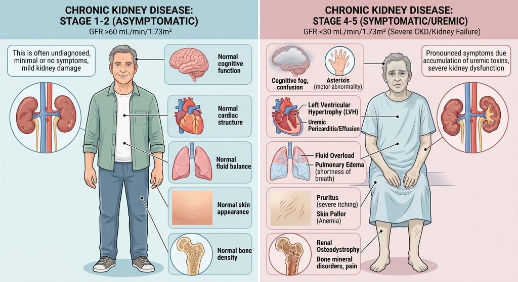
Symptoms are described by the patient, while signs are objective clinical findings discovered by the medical doctor. The insidious nature of Chronic Kidney Disease stems from the kidney’s capacity to adapt through single-nephron hyperfiltration. Patients can lose up to 70% of their functional nephron mass without overt clinical symptoms. When signs appear in stages G4 and G5, they reflect multi-organ system toxicity:
| Sign or Symptom | Clinical Presentation | Pathophysiological Mechanism |
| Peripheral and Periorbital Edema | Pitting edema of the lower extremities, morning periorbital puffiness, ascites | Loss of GFR impairs fractional sodium and water clearance; nephrotic-range proteinuria causes hypoalbuminemia, reducing plasma oncotic pressure. |
| Asthenia, Pallor, and Fatigue | Profound, unremitting exhaustion, exercise intolerance, pale conjunctivae | Deficiency of renal peritubular erythropoietin production combined with hepcidin-mediated iron trapping causes normocytic anemia. |
| Hypertension (Worsening) | Treatment-resistant elevated blood pressure, morning occipital headaches | Intravascular volume expansion from impaired sodium excretion, uninhibited systemic RAAS activity, and elevated sympathetic nervous tone. |
| Nocturia and Polyuria | Waking 2–4 times per night to void, passing dilute urine | Disruption of the hypertonic medullary countercurrent gradient impairs tubular responsiveness to ADH (vasopressin), reducing urinary concentrating ability. |
| Dyspnea and Orthopnea | Shortness of breath, bibasilar crackles on auscultation, intolerance to lying flat | Hydrostatic pulmonary capillary transudation from fluid overload; compensatory tachypneic hyperventilation (Kussmaul breathing) for metabolic acidosis. |
| Nausea, Vomiting, Dysgeusia | Morning retching, early satiety, metallic taste, uremic fetor (ammoniacal breath) | Retained middle molecules and nitrogenous wastes irritate gastric mucosa and stimulate the medullary chemoreceptor trigger zone; oral bacteria convert urea to ammonia. |
| Severe Pruritus | Intractable, diffuse skin itching without primary rash, excoriations | Dermal micro-precipitation of calcium-phosphate crystals, secondary hyperparathyroidism, peripheral sensory neuropathy, and uremic neurotoxins. |
| Restless Legs and Asterixis | Involuntary leg movements at night, bilateral flapping tremor of dorsiflexed hands | Neurotoxicity driven by guanidino compounds, parathyroid hormone elevation, metabolic encephalopathy, and microvascular central nervous ischemia. |
| Uremic Pericarditis | Sharp pleuritic chest pain relieved by sitting forward, pericardial friction rub | Direct inflammatory chemical irritation of visceral and parietal pericardial layers by uremic serosal exudate; indication for emergent dialysis. |
| Bone Pain and Fragility Fractures | Deep skeletal pain in hips, spine, and ribs; low-trauma stress fractures | Renal osteodystrophy caused by hyperphosphatemia, low 1,25(OH)2D, secondary hyperparathyroidism, and osteoclastic bone resorption. |
6. Chronic Kidney Disease Risk Factors
Section Summary:
Risk factors for CKD are divided into initiation factors, which trigger primary renal parenchymal damage, and progression factors, which accelerate the loss of remaining nephrons. While genetics and age are fixed, managing modifiable risks—like hypertension, glycemic control, and nephrotoxic drug exposures—can alter disease trajectory.
Identifying risk factors enables early screening, targeted intervention, and risk mitigation. Risk factors are divided into modifiable and non-modifiable:
- Type 2 Diabetes Mellitus: a modifiable risk factor. Hyperglycemia drives advanced glycation end products, efferent arteriolar constriction, and mesangial expansion. For a better understanding, please read our Type 2 Diabetes Mellitus article.
- Systemic Hypertension: a modifiable risk factor. Chronic barotrauma destroys glomerular capillary loops, accelerating glomerulosclerosis. For a better understanding, please read our Hypertension article.
- Cardiovascular Disease: a modifiable risk factor. Low cardiac output or venous congestion alters renal perfusion, triggering cardiorenal syndrome.
- Persistent Proteinuria: a modifiable risk factor. Intraluminal albumin causes tubular toxicity, peritubular inflammation, and interstitial fibrosis.
- Nephrotoxic Medications: a modifiable risk factor. Chronic Nonsteroidal Antinflammatory Drugs (NSAID) use inhibits renal prostacyclin, causing ischemic medullary necrosis; aminoglycosides and contrast media cause tubular toxicity.
- Cigarette Smoking: a modifiable risk factor. Nicotine stimulates sympathetic outflow and endothelin-1, accelerating endothelial dysfunction and glomerulosclerosis.
- Excess Weight (BMI ≥ 30): a modifiable risk factor. Hyperfiltration nephropathy and increased metabolic demands cause compensatory glomerular enlargement and secondary FSGS.
- Advanced Age (>60 years): a non-modifiable risk factor. Age-related glomerulosclerosis causes a physiological eGFR decline of ~0.75–1.0 mL/min/1.73 m² per year after age 40.
- Family History / Genetics: a non-modifiable risk factor. Genetic variants (APOL1 high-risk alleles, PKD1, PKD2, COL4A3/4/5) increase susceptibility to glomerular and cystic disease.
- Prior Episode of Acute Kidney Injury: a non-modifiable risk factor. Acute tubular necrosis can leave residual fibrosis, microvascular rarefaction, and reduced nephron reserve.
7. Chronic Kidney Disease Diagnostic Criteria
Section Summary:
A definitive diagnosis of Chronic Kidney Disease requires laboratory verification of reduced eGFR (< 60 mL/min/1.73 m²) or structural/functional renal damage persisting for more than three months. Standard diagnostic workup involves the CKD-EPI 2021 race-free creatinine-cystatin C equation, a spot urine Albumin-to-Creatinine Ratio (uACR), urinalysis with sediment microscopy, and renal ultrasonography.
This section will begin with the algorithm for diagnosing Chronic Kidney Disease:
Step 1: Patient Identification & Screening Indications
Evaluate patients presenting with established risk factors, including:
- Type 1 or Type 2 Diabetes Mellitus
- Systemic Hypertension
- Established Cardiovascular Disease (Heart Failure, Coronary Artery Disease, Stroke)
- Family history of kidney disease or genetic renal disorders
- Previous history of Acute Kidney Injury (AKI)
- Age > 60 years, persistent hematuria, or incidental abnormal laboratory findings
Step 2: Initial Dual-Marker Laboratory Screening
Obtain both filtration and structural/barrier integrity markers simultaneously:
- Serum Creatinine & Cystatin C: Calculate estimated Glomerular Filtration Rate (eGFR) using the race-free CKD-EPI 2021 equation.
- Spot Urine Sample: Collect a first-morning void to measure the Urine Albumin-to-Creatinine Ratio (uACR).
- Dipstick Urinalysis: Screen for hematuria, pyuria, and proteinuria.
Step 3: Threshold Evaluation
Assess whether either core diagnostic threshold is met:
- Filtration Impairment: eGFR (< 60 mL/min/1.73 m²).
- Kidney Damage Marker: uACR ≥ 30 mg/g (≥ 3 mg/mmol), persistent microscopic hematuria of glomerular origin, or structural defects on imaging
Decision Branch:
- Neither threshold is met: Chronic Kidney Disease is excluded. Continue routine annual surveillance for high-risk patients (e.g., individuals with diabetes or hypertension).
- Either threshold is met: Proceed to Step 4 to establish chronicity.
Step 4: Chronicity Assessment (The 3-Month Rule) Determine whether the renal abnormality represents an acute process (AKI/AKD) or chronic parenchymal loss:
- Review Prior Medical Records: Compare current results with historical serum creatinine, eGFR, or uACR values from at least 3 months prior.
- Assess Structural Chronicity: Perform a renal ultrasound. Findings of bilateral cortical thinning (< 1 cm), reduced renal length (< 9 cm), or increased echogenicity support chronicity.
Step 5: Diagnostic Confirmation & Clinical Pathway
Pathway A — CKD Confirmed (Abnormality Documented for > 3 Months):
- Establish the underlying etiology (diabetic nephropathy, hypertensive nephrosclerosis, glomerulonephritis, cystic disease).
- Assign the KDIGO CGA stage (Cause, GFR category G1–G5, Albuminuria category A1–A3).
- Initiate guideline-directed organ-protective therapy (RAAS inhibitors, SGLT2 inhibitors, non-steroidal MRAs, blood pressure control).
- Refer to nephrology if eGFR < 30 mL/min/1.73 m², uACR > 300 mg/g, or if rapid functional decline is detected.
Pathway B — Unconfirmed Chronicity (No Historical Data Available):
- Classify provisionally as an Acute Kidney Disease (AKD) or investigate for resolving/active Acute Kidney Injury (AKI).
- Discontinue potential nephrotoxins (e.g., NSAIDs, aminoglycosides) and correct reversible hemodynamic factors.
- Re-test eGFR, uACR, and urinalysis at 3 months. If abnormalities persist at 3 months → Confirm Chronic Kidney Disease diagnosis (follow Pathway A). If abnormalities resolve → Document resolved AKI/AKD and schedule periodic re-evaluation.
Because CKD is typically asymptomatic in its early stages, diagnosis requires systematic screening of at-risk populations:
1. Verification of the 3-Month Chronicity Window
A single abnormal laboratory value cannot establish a diagnosis of CKD. An isolated elevated creatinine or depressed eGFR may reflect Acute Kidney Injury (prerenal azotemia, acute tubular necrosis, or drug toxicity).
Chronicity is established by:
- Reviewing prior historical serum creatinine, eGFR, or urinalysis results spanning >3 months.
- If historical data are absent, repeat testing in 3 months.
- Demonstrating structural changes on renal imaging (e.g., small, shrunken kidneys or advanced cystic disease) that indicate long-standing injury.
2. Assessment of Glomerular Filtration Rate (eGFR)
The gold standard for measuring GFR (inulin or iohexol clearance) is clinically impractical. Therefore, validated serum biomarkers and estimation equations are used:
- Serum Creatinine: Creatinine is generated by muscle metabolism and cleared by glomerular filtration. However, its concentration is influenced by muscle mass, dietary meat intake, age, sex, and tubular secretion (10–15%).
- Serum Cystatin C: A low-molecular-weight protein produced by all nucleated cells at a constant rate, freely filtered by the glomerulus, and metabolized by proximal tubular cells. Cystatin C is unaffected by muscle mass, dietary protein, or acute illness.
- The CKD-EPI 2021 Equations: The KDIGO 2024 guidelines recommend the race-free CKD-EPI 2021 equations. When higher precision is required—such as confirming an eGFR between 45–59 mL/min/1.73 m² without albuminuria, or in patients with abnormal muscle mass—the combined creatinine-cystatin C equation eGFR (cr-cys).
3. Quantification of Albuminuria: Spot Urine uACR
The 24-hour urine collection has been largely replaced in routine practice due to collection errors and patient burden. KDIGO recommends the urine Albumin-to-Creatinine Ratio (uACR) on a random first-morning spot urine sample:
- Why Albumin, Not Total Protein? Albumin is the most sensitive marker for glomerular filtration barrier damage. Total protein dipsticks are insensitive to early microalbuminuria and primarily detect albumin only when levels exceed 300 mg/day.
- Creatinine Correction: Measuring the ratio of urinary albumin (mg/dL) to urinary creatinine (g/dL) controls for fluctuations in urine concentration driven by hydration status.
- Confirmation Rule: Because transient albuminuria can be induced by strenuous exercise, fever, urinary tract infection, decompensated heart failure, or upright posture (orthostatic proteinuria), a diagnosis of persistent albuminuria requires at least two of three abnormal uACR tests collected over a 3- to 6-month period.
4. Urinalysis and Sediment Examination
Urine microscopy provides direct cellular clues about the primary injury mechanism:
- Dysmorphic Erythrocytes & Acanthocytes (G1 cells): Erythrocytes deformed by passage through an injured basement membrane indicate active glomerulonephritis.
- Cellular Casts. Red Blood Cell Casts: Pathognomonic for active glomerulonephritis or vasculitis. White Blood Cell Casts: Indicative of tubulointerstitial nephritis or pyelonephritis. Broad, Waxy Casts: Formed in dilated, atrophic tubules; indicative of advanced end-stage renal disease.
- Sterile Pyuria: White blood cells in the absence of bacterial growth suggest tubulointerstitial nephritis or renal tuberculosis.
5. Renal Ultrasonography
Renal ultrasound is a non-invasive imaging modality that helps establish chronicity, evaluate gross anatomy, and rule out obstruction:
- Kidney Size: Normal renal bipolar length is 10 to 12 cm. Bilateral small kidneys (<9 cm) indicate chronic parenchymal atrophy and irreversible fibrosis.
- Cortical Thickness: Normal renal cortex is ≥ 1 cm. Cortical thinning and increased cortical echogenicity (brighter than normal liver parenchyma) indicate diffuse glomerulosclerosis and interstitial fibrosis.
- Exceptions to Small Kidneys in Chronic Kidney Disease: Normal or enlarged kidneys in advanced Chronic Kidney Disease occur in Diabetic Kidney Disease (early to mid-stages), Amyloidosis, HIV-associated nephropathy, and Autosomal Dominant Polycystic Kidney Disease.
- Hydronephrosis: Dilation of the renal pelvicalyceal system indicates post-renal obstructive nephropathy.
6. Indications for Percutaneous Renal Biopsy
Renal biopsy provides direct histological characterization but carries a small risk of bleeding. Biopsy is indicated when non-invasive testing fails to establish an etiology or when findings will directly guide therapy:
- Unexplained acute decline in GFR or rapid progression of Chronic Kidney Disease.
- Nephrotic-range proteinuria (uACR > 2200 mg/g or protein > 3.5 g/24h) without a clear systemic cause.
- Active urinary sediment (dysmorphic hematuria, cellular casts) suggests rapidly progressive glomerulonephritis or vasculitis.
- Renal involvement in systemic autoimmune diseases (e.g., suspected class III–V Lupus Nephritis).
8. Chronic Kidney Disease Treatment and Prevention
Section Summary:
Modern Chronic Kidney Disease management focuses on disease modification via four foundational pharmacotherapies: maximum tolerated RAAS inhibition, SGLT2 inhibitors, non-steroidal mineralocorticoid receptor antagonists, and GLP-1 receptor agonists. This is combined with strict blood pressure control (systolic < 120 mmHg), statin-based cardiovascular risk management, and dietary optimization.

The therapeutic approach to CKD has transitioned from supportive care to active disease modification:
Foundational Pharmacotherapy (The Cardiorenal Pillars)
| Drug Class | Examples | Target Population | Mechanism of Action | Clinical Trial Evidence |
| ACE Inhibitors / ARBs | Lisinopril, Ramipril / Losartan, Enalapril | All patients with hypertension, CKD, and uACR ≥ 30–300 mg/g | Selectively dilates efferent arterioles, reducing intraglomerular hypertension and mechanical shear stress; suppresses angiotensin-driven fibrosis. | RENAAL, IDNT, RIT trials: Reductions in proteinuria and progression to ESRD. Titrated to maximum tolerated dose. |
| SGLT2 Inhibitors | Dapagliflozin, Empagliflozin | Patients with CKD with eGFR ≥ 20 mL/min/1.73 m², with or without Type 2 Diabetes | Blocks proximal tubular glucose and sodium reabsorption, restoring tubuloglomerular feedback (macula densa sodium sensing) to constrict afferent arterioles and reduce intraglomerular pressure. | DAPA-CKD, EMPA-KIDNEY: 28–39% relative risk reduction in CKD progression, ESRD, and cardiovascular death across diabetic and non-diabetic cohorts. |
| Non-Steroidal MRAs | Finerenone | CKD associated with Type 2 Diabetes with persistent albuminuria despite maximally tolerated RAAS blockade | Selectively blocks mineralocorticoid receptor overactivation with minimal impact on blood pressure, suppressing downstream pro-inflammatory and pro-fibrotic signaling. | FIDELIO-DKD, FIGARO-DKD: Significant reductions in composite kidney failure and cardiovascular events; lower hyperkalemia risk than spironolactone. |
| GLP-1 Receptor Agonists | Semaglutide, Dulaglutide | CKD with Type 2 Diabetes, particularly with obesity or established atherosclerotic cardiovascular disease | Incretin mimetic: reduces body weight, improves glycemic control, attenuates systemic inflammation, and reduces renal vascular resistance. | FLOW Trial: Halted early due to demonstrated efficacy in reducing major kidney outcomes and cardiovascular death in diabetic kidney disease. |
Clinical Note on Hemodynamic eGFR Dips: Initiating an ACEi, ARB, or SGLT2 inhibitor frequently causes an acute, benign decline in eGFR of up to 30% from baseline. This reflects the desired reduction in intraglomerular hyperfiltration. These medications should not be discontinued unless the eGFR drop exceeds 30% or severe, refractory hyperkalemia occurs.
Blood Pressure and Cardiovascular Risk Optimization
- Standardized Blood Pressure Targets: The KDIGO 2024 guidelines recommend targeting a systolic blood pressure < 120 mmHg, measured using standardized office techniques, when tolerated. This intensive target slows GFR decline and reduces cardiovascular mortality, though blood pressure targets should be individualized for frail patients.
- Lipid Management: Patients with Chronic Kidney Disease are at high risk for atherosclerotic events. KDIGO recommends statin-based therapy (e.g., Atorvastatin 20 mg daily or combined Simvastatin/Ezetimibe) for all adults aged ≥ 50 years with Chronic Kidney Disease non-dialysis dependent, regardless of baseline LDL-cholesterol.
Management of Metabolic Complications
- Renal Anemia: Maintain hemoglobin between 10.0 and 11.5 g/dL. Correct iron deficiency first, targeting a ferritin > 100 ng/mL and transferrin saturation (TSAT) > 20% using oral or intravenous iron. If anemia persists, initiate Erythropoiesis-Stimulating Agents (ESAs) (e.g., Darbepoetin alfa, Epoetin alfa) or oral HIF-Prolyl Hydroxylase Inhibitors (HIF-PHIs) (e.g., Roxadustat).
- CKD-MBD: Control serum phosphorus toward normal (2.5–4.5 mg/dL) using dietary restriction and intestinal phosphate binders. Calcium-free binders: Sevelamer carbonate, Lanthanum carbonate (preferred to prevent vascular calcification). Calcium-based binders: Calcium acetate (use with caution to avoid calcium overload). Vitamin D Therapy: For secondary hyperparathyroidism, use nutritional vitamin D (cholecalciferol) or active vitamin D analogs (Calcitriol, Paricalcitol) to suppress elevated intact PTH.
- Metabolic Acidosis: Treat serum bicarbonate concentrations < 20–22 mEq/L with oral Sodium Bicarbonate (0.5 to 1.0 mEq/kg/day) to prevent accelerated muscle proteolysis and slow GFR decline.
- Hyperkalemia Management: Avoid unnecessary discontinuation of RAAS inhibitors due to hyperkalemia. Instead, optimize dietary potassium intake and utilize modern potassium-binding polymers: Patiromer or Sodium Zirconium Cyclosilicate (SZC).
Preparation for Kidney Replacement Therapy (KRT)
When a patient progresses to late Stage G4 or early Stage G5 (or a 5-year kidney failure risk > 3–5 by the Kidney Failure Risk Equation), education regarding kidney replacement therapy options must begin:
- Preemptive Kidney Transplantation: The preferred therapy for eligible candidates, providing superior long-term survival and quality of life compared to dialysis.
- Hemodialysis (HD) requires planning and early creation of an arteriovenous (AV) fistula in the non-dominant arm at least 3 to 6 months prior to anticipated dialysis initiation, avoiding central venous catheters.
- Peritoneal Dialysis (PD): An option for home-based therapy utilizing the peritoneal membrane as a biological filter.
- Comprehensive Conservative Kidney Management: A patient-centered, non-dialytic pathway focused on symptom control, advance care planning, and quality-of-life preservation.
9. Chronic Kidney Disease Prognosis and Interconnection with Other Diseases
Section Summary:
Patients with Chronic Kidney Disease are significantly more likely to die from cardiovascular disease than to progress to end-stage renal disease. The condition is closely linked to Type 2 Diabetes, Hypertension, Congestive Heart Failure, Coronary Heart Disease, and Atrial Fibrillation via shared mechanisms within the cardiorenal-metabolic syndrome.
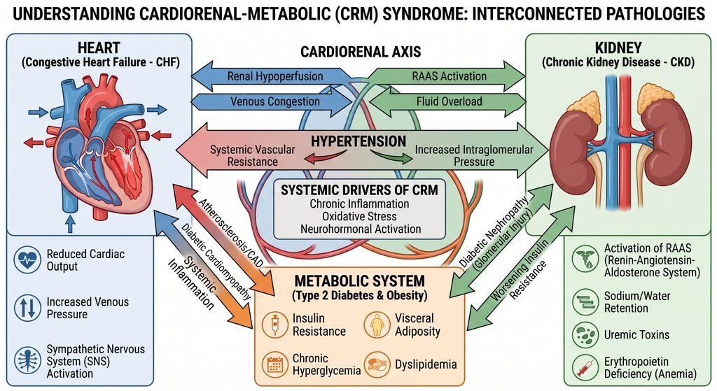
The prognosis of Chronic Kidney Disease is largely driven by its systemic complications. Rather than progressing to dialysis, the leading cause of death in Chronic Kidney Disease across all stages is cardiovascular disease. Chronic Kidney Disease interacts directly with other common cardiometabolic conditions:
1. Connection with Type 2 Diabetes Mellitus
Diabetic Kidney Disease (DKD) is the primary driver of end-stage renal disease globally. Chronic hyperglycemia activates protein kinase C, increases advanced glycation end-products, and causes sorbitol accumulation. In early stages, this induces glomerular hyperfiltration; in later stages, it leads to basement membrane thickening, nodular glomerulosclerosis, and nephron loss.
Conversely, progressive renal failure disrupts normal insulin clearance, resulting in unpredictable hypoglycemia in patients maintained on fixed doses of insulin or insulin secretagogues. For a complete understanding, please read our Type 2 Diabetes Mellitus article.
2. Connection with Hypertension
Hypertension serves as both a primary cause and an inevitable consequence of Chronic Kidney Disease. Uncontrolled systemic pressures damage renal arterioles, leading to nephrosclerosis.
As functioning nephron mass declines, impaired natriuresis leads to intravascular volume expansion. Coupled with persistent renin secretion, this elevates systemic vascular resistance and blood pressure, creating a progressive cycle of vascular and renal injury. For a better understanding, please read our Hypertension article.
3. Connection with Congestive Heart Failure (The Cardiorenal Syndrome)
Chronic Kidney Disease and Heart Failure frequently coexist within the framework of Cardiorenal Syndrome (Types 2 and 4):
- Impaired cardiac output decreases renal perfusion, activating neurohormonal pathways (RAAS and the sympathetic nervous system) that promote renal sodium and water retention.
- Venous congestion increases central venous pressure, which is transmitted to the renal veins, lowering the transglomerular pressure gradient and accelerating GFR decline.
- Chronic kidney disease accelerates left ventricular hypertrophy, myocardial interstitial fibrosis, and volume overload, contributing to both heart failure with preserved ejection fraction (HFpEF) and heart failure with reduced ejection fraction (HFrEF).
For a complete clinical guide, please read our Congestive Heart Failure article.
4. Connection with Coronary Heart Disease (CAD)
Atherosclerosis in Chronic Kidney Disease involves distinct pathophysiological mechanisms compared to the general population:
- In addition to classic intimal lipid plaques, Chronic Kidney Disease patients develop extensive Mönckeberg medial calcification driven by hyperphosphatemia, hyperparathyroidism, and elevated FGF-23.
- This extensive medial calcification causes arterial stiffness, widened pulse pressure, and impaired subendocardial coronary perfusion, markedly increasing the incidence of non-ST-elevation myocardial infarction and sudden cardiac death.
For a complete understanding, please read our Coronary Artery Disease article.
5. Connection with Atrial Fibrillation (AFib)
Chronic volume overload elevates left ventricular filling pressures, causing progressive left atrial enlargement and fibrosis. Coupled with electrolyte shifts (hyperkalemia, hypocalcemia) and systemic uremic inflammation, this markedly increases the incidence of Atrial Fibrillation.
Managing AFib in Chronic Kidney Disease requires careful risk assessment: impaired renal clearance increases the bleeding risk of direct oral anticoagulants (DOACs), so dose adjustments should be based on creatinine clearance. To better understand this condition, please read our Atrial Fibrillation article.
6. Correlation with Routine Blood Tests
Active surveillance of routine clinical blood tests is essential in managing the systemic complications of Chronic Kidney Disease:
- Serum Creatinine & BUN: Primary markers for tracking clearance and calculating eGFR.
- Electrolyte Panel (K+, Na+, HCO3-): Identifies life-threatening hyperkalemia and subclinical metabolic acidosis.
- Serum Calcium, Phosphate, and Intact PTH: Monitored to detect early mineral and bone disorders (CKD-MBD).
- Complete Blood Count (CBC): Tracks the development and progression of normocytic renal anemia.
For a complete guide to every value, please read our Blood Test article.
10. Living with Chronic Kidney Disease
Section Summary:
Daily management of Chronic Kidney Disease centers on nutritional adjustments, cardiovascular risk reduction, and protecting remaining renal function. Core measures include following a low-sodium, plant-dominant diet, staying physically active, understanding acute “sick-day rules” to avoid medication-induced nephrotoxicity, and taking steps to preserve vascular access.
There are various daily life modifications that are mandatory to be implemented in patients with Chronic Kidney Disease.
Dietary Sodium Restriction: Limit dietary sodium to < 2.0 g of sodium per day (< 5.0 g of sodium chloride/table salt). Restricting sodium lowers blood pressure, reduces fluid retention, and enhances the antiproteinuric effects of RAAS inhibitors.
Dietary Protein Management:
- Stages G3–G5 (non-dialysis): A moderate protein intake of 0.6 – 0.8 g/kg/day is recommended to reduce nitrogenous waste and slow renal functional decline while preventing protein-energy wasting.
- Dialysis (G5D): Protein targets increase to 1.0 – 1.2 g/kg/day to compensate for dialysate amino acid losses and chronic catabolism.
Potassium and Phosphorus Management: In stages G4–G5, elevated serum phosphorus requires limiting high-phosphate foods (processed foods with phosphate additives, dark colas, processed meats). Dietary potassium should be tailored based on serum levels rather than restricted preemptively, encouraging nutrient-dense, plant-rich dietary patterns.
The “SADMANS” Sick-Day Rules: During acute intercurrent illnesses causing dehydration (e.g., gastroenteritis, vomiting, fever), patients should know which medications to temporarily pause to prevent acute-on-chronic kidney injury:
- S: Sulfonylureas (prevents severe hypoglycemia)
- A: ACE inhibitors (prevents abrupt drop in glomerular filtration)
- D: Diuretics (prevents severe hypovolemia)
- M: Metformin (prevents lactic acidosis)
- A: Angiotensin Receptor Blockers (ARBs)
- N: NSAIDs (strictly avoided due to afferent vasoconstriction)
- S: SGLT2 inhibitors (prevents euglycemic DKA and volume depletion)
- Vascular Access Preservation (“Save the Veins”): For individuals with Stage G4 or G5 Chronic Kidney Disease, preserving the cephalic and basilic veins in the non-dominant arm is essential for future AV fistula creation. Peripheral intravenous lines, venipunctures, and PICC lines should be placed in the dominant arm or dorsum of the hand whenever possible.
11. Chronic Kidney Disease Frequently Asked Questions (FAQs)
Section Summary:
This section provides concise, evidence-based answers to common patient questions regarding the reversibility of Chronic Kidney Disease, progression timelines, dietary considerations, hydration needs, and the silent nature of early-stage disease.

Can Chronic Kidney Disease be cured or reversed?
Established structural kidney damage and fibrosis in Chronic Kidney Disease are irreversible. However, the disease process can often be stabilized. With modern quadruple therapy (RAAS inhibitors, SGLT2 inhibitors, non-steroidal MRAs, and GLP-1 RAs), blood pressure control, and lifestyle interventions, the rate of eGFR decline can be substantially slowed, allowing many patients to avoid end-stage renal disease.
What is the difference between Acute Kidney Injury and Chronic Kidney Disease?
Acute Kidney Injury (AKI) is an abrupt decline in kidney function occurring over hours to days, which is often reversible once the underlying trigger (e.g., dehydration, sepsis, toxic medication) is corrected. Chronic Kidney Disease (CKD) represents progressive structural and functional damage persisting for greater than 3 months. However, an episode of AKI can accelerate underlying CKD, and CKD is itself a major risk factor for AKI.
How long can someone remain in Stage 3 Chronic Kidney Disease without progressing to dialysis?
Many patients remain in Stage 3 (G3a/G3b) for years or decades, and some never require dialysis. Progression depends heavily on the level of albuminuria, blood pressure management, glycemic control, and medication adherence. Patients with low albuminuria (A1) have a low lifetime risk of kidney failure compared to those with severe albuminuria (A3).
Should people with Chronic Kidney Disease drink large amounts of water to “flush out toxins”?
Drinking excess water does not improve renal clearance and will not reverse kidney disease. In early stages (G1–G2), drinking to thirst is generally appropriate. In advanced stages (G4–G5), failing kidneys lose the ability to clear free water; excessive fluid intake can cause dilution hyponatremia, peripheral edema, hypertension, and pulmonary edema. Fluid intake should be individualized with guidance from a healthcare provider.
Why does Chronic Kidney Disease cause no pain or warning signs in its early stages?
Renal parenchymal tissue lacks sensory pain fibers; nociceptors are located primarily in the renal capsule and ureteric system. Pain typically occurs only when the capsule is rapidly stretched (as in acute pyelonephritis, renal cyst rupture, or acute obstruction) or when a stone passes down the ureter. Because chronic nephron loss occurs gradually without stretching the capsule, Chronic Kidney Disease is painless and clinically silent until advanced uremic toxicity develops.
12. High-Yield Chronic Kidney Disease Questions to Ask Your Doctor
Section Summary:
To support patient advocacy and clinical communication, this section outlines essential questions for patients to ask their healthcare provider regarding their eGFR trajectory, albuminuria levels, medication management, and specialized nephrology care.

“What is my current eGFR, what is my urine albumin-to-creatinine ratio (uACR), and how have they changed over time?”
- Clinical Rationale: Understanding both values helps define your overall risk category under KDIGO guidelines and track your rate of functional decline.
“Am I taking medications that protect my kidneys, such as an ACE inhibitor, ARB, or SGLT2 inhibitor?”
- Clinical Rationale: These drug classes slow disease progression in eligible patients and reduce cardiorenal complications.
“Are any of my current prescription or over-the-counter medications potentially nephrotoxic?”
- Clinical Rationale: Identifying common nephrotoxic agents—such as NSAIDs, proton pump inhibitors, and certain antibiotics—helps protect remaining nephrons.
“What is my individualized blood pressure target, and should I track my readings at home?”
- Clinical Rationale: While guidelines often target a systolic reading below 120 mmHg, personal goals must account for age, frailty, and cardiovascular status.
“Do my lab results or disease stage warrant a formal referral to a nephrologist or a renal dietitian?”
- Clinical Rationale: Early nephrology referral (typically when eGFR drops below 30 mL/min/1.73 m² or if significant albuminuria/hematuria is present) improves outcomes and allows timely education on kidney replacement options.
“What should I do with my medications if I become ill with vomiting, diarrhea, or fever?”
- Clinical Rationale: Knowing when to temporarily pause specific medications (the “SADMANS” rules) reduces the risk of acute-on-chronic kidney injury.
Credible Sources and References
- Kidney Disease: Improving Global Outcomes (KDIGO) CKD Work Group. KDIGO 2024 Clinical Practice Guideline for the Evaluation and Management of Chronic Kidney Disease. Kidney International, 2024; 105(4S): S117–S314.
- American Diabetes Association (ADA). Standards of Care in Diabetes—2026. Chronic Kidney Disease and Risk Management. Diabetes Care, 2026; 49(Suppl. 1).
- World Health Organization (WHO). Global Health Estimates: Noncommunicable Diseases and Chronic Kidney Disease Burden, Geneva: WHO Press.
- Heerspink HJL, Stefánsson BV, Correa-Rotter R, et al. Dapagliflozin in Patients with Chronic Kidney Disease (DAPA-CKD). New England Journal of Medicine, 2020; 383(15): 1436–1446.
- The EMPA-KIDNEY Collaborative Group. Empagliflozin in Patients with Chronic Kidney Disease. New England Journal of Medicine, 2023; 388(2): 117–127.
- Bakris GL, Agarwal R, Anker SD, et al. Effect of Finerenone on Chronic Kidney Disease Outcomes in Type 2 Diabetes (FIDELIO-DKD). New England Journal of Medicine, 2020; 383(23): 2219–2229.
- Perkovic V, Tuttle KR, Rossing P, et al. Effects of Semaglutide on Chronic Kidney Disease Outcomes in Patients with Type 2 Diabetes (FLOW Trial). New England Journal of Medicine, 2024; 391(2): 109–121.
- Inker LA, Eneanya ND, Coresh J, et al. New Creatinine- and Cystatin C–Based Equations to Estimate GFR without Race. New England Journal of Medicine, 2021; 385(19): 1737–1749.
- National Kidney Foundation. KDOQI Clinical Practice Guideline for Hemodialysis Adequacy and Vascular Access Updates. American Journal of Kidney Diseases.
Featured Image: Conceptual 3D medical rendering illustrating the internal macro- and microvascular filtration architecture of the human kidney, created with the help of Artificial Intelligence.
© UncutMed — Medical knowledge, precise, truthful, unfiltered. The current article was last modified on September 16, 2026. This article was clinically authored and reviewed under international evidence-based standards. Always consult your personal healthcare physician before modifying any medical therapy.