Copyright UncutMed. Medical knowledge truthful, precise, unfiltered. This article was last reviewed on September 3, 2026. Always consult your healthcare provider for personal medical advice.
Medical Disclaimer: This article is for informational and educational purposes only. It does not constitute medical advice, diagnosis, or treatment. Always consult a qualified healthcare professional regarding any medical condition or treatment plan.
Atrial Fibrillation – Medical Definition
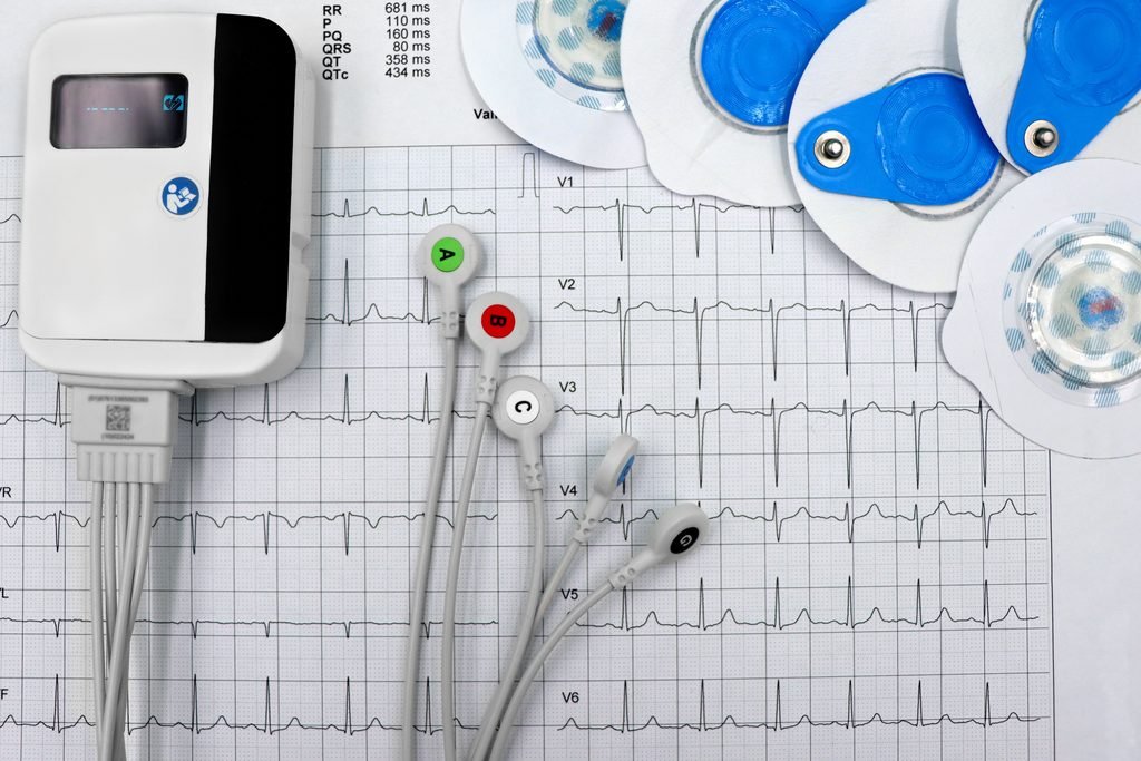
Atrial fibrillation (AFib) is a supraventricular tachyarrhythmia characterised by uncoordinated atrial electrical activation with consequent deterioration of atrial mechanical function. It is defined electrocardiographically by the replacement of consistent P waves by rapid oscillatory or fibrillatory waves that vary in amplitude, shape, and timing, associated with an irregular, frequently rapid ventricular response when atrioventricular conduction is intact.
In physiological terms, Atrial Fibrillation represents a state of chaotic, disorganised electrical activity within the atrial myocardium. Rather than a single coordinated electrical impulse originating from the sinoatrial (SA) node propagating in an orderly fashion through the atria to the atrioventricular (AV) node, Atrial Fibrillation is driven by multiple simultaneous wavefronts of electrical activity circulating through the atrial tissue in a disorganised, self-perpetuating manner. The atria quiver at rates between 350 and 600 impulses per minute, rendering them haemodynamically ineffective.
The Atrioventricular Node (AV) filters this torrential bombardment of atrial impulses, allowing only a fraction to conduct to the ventricles. The result is the clinically characteristic irregularly irregular ventricular rhythm — the defining feature of AF on both electrocardiography and pulse palpation.
Atrial Fibrillation is the most common sustained cardiac arrhythmia in clinical practice, affecting an estimated 37.5 million people worldwide. Its prevalence increases dramatically with age — affecting approximately 0.5% of adults aged 40-50, rising to over 10% in those aged 80 and above. It is associated with a fivefold increased risk of ischaemic stroke, a threefold increased risk of heart failure, and a twofold increased risk of all-cause mortality compared to age-matched individuals in sinus rhythm.
Atrial Fibrillation – Classification
AF is classified into five clinical subtypes:
- First Detected Atrial Fibrillation — the first diagnosed episode, regardless of whether it is symptomatic or self-terminating.
- Paroxysmal Atrial Fibrillation — episodes that terminate spontaneously or with intervention within 7 days of onset.
- Persistent Atrial Fibrillation — AF that fails to terminate spontaneously and lasts more than 7 days, or is terminated by pharmacological or electrical cardioversion.
- Long-Standing Persistent Atrial Fibrillation — continuous AF lasting more than 12 months when a rhythm control strategy is pursued.
- Permanent Atrial Fibrillation — AF in which the presence of the arrhythmia is accepted by the patient and clinician, with no further attempts at rhythm restoration.
Atrial Fibrillation – Pathophysiology
The pathophysiology of Atrial Fibrillation is complex and multifactorial, involving the interaction of triggers, an arrhythmogenic substrate, and modulatory influences — most notably autonomic nervous system activity.
Triggers of Atrial Fibrillation Initiation
The most important and well-characterised triggers of Atrial Fibrillation initiation are ectopic electrical foci located within the pulmonary veins (PVs). This is the mechanism that initiates the episodes of Atrial Fibrillation. The myocardial sleeves extending from the left atrium into the proximal pulmonary veins possess unique electrophysiological properties — including abnormal automaticity, triggered activity, and the capacity for micro-reentry — that predispose them to spontaneous ectopic firing. These PV ectopic beats fall on vulnerable periods of the atrial myocardium’s repolarisation cycle, triggering Atrial Fibrillation initiation in susceptible individuals.
The cardinal importance of pulmonary vein triggers is the electrophysiological basis for pulmonary vein isolation (PVI) — the cornerstone of catheter ablation therapy for Atrial Fibrillation.
Beyond the pulmonary veins, non-PV triggers — including the superior vena cava, the ligament of Marshall, the crista terminalis, the coronary sinus, and the posterior left atrial wall — may contribute to AF initiation in a subset of patients, particularly those with persistent or long-standing persistent Atrial Fibrillation.
The Arrhythmogenic Substrate
For Atrial Fibrillation to be maintained beyond its initial trigger, an arrhythmogenic substrate must exist within the atrial myocardium.
Atrial Fibrosis: The most important substrate abnormality in Atrial Fibrillation. This is the replacement of normal atrial myocardium with collagen-rich fibrous tissue. Fibrosis disrupts normal electrical coupling between adjacent myocardial cells, creating areas of slow conduction, unidirectional block, and heterogeneous refractoriness — ideal conditions for the initiation and perpetuation of re-entrant circuits. Atrial fibrosis results from hypertension (for more information, please read our respective article), heart failure (for more information, please read our respective article), ageing, inflammation, and obesity.
Electrical Remodelling: Sustained rapid atrial activation in Atrial Fibrillation produces progressive changes in atrial ion channel expression — particularly downregulation of the L-type calcium current (ICaL) — which shorten atrial action potential duration and effective refractory period. This electrical remodelling reduces the wavelength of re-entrant circuits, allowing more simultaneous wavefronts to coexist within the atria and stabilising Atrial Fibrillation. This phenomenon underpins the well-established clinical aphorism “AF begets AF.”
Atrial Dilatation: Increased atrial chamber size provides greater total atrial mass for re-entrant wavefronts to circulate, facilitating Atrial Fibrillation perpetuation. Atrial dilatation is both a cause and consequence of AFib, creating a self-reinforcing cycle.
Autonomic Modulation
Both heightened vagal tone — which shortens atrial refractoriness — and heightened sympathetic tone — which increases automaticity and triggered activity — can facilitate Atrial Fibrillation initiation. This explains why AFib can be triggered by both intense physical exertion and by vagal manoeuvres or sleep.
Haemodynamic Consequences of Atrial Fibrillation
Loss of Atrial Systole: In normal sinus rhythm, coordinated atrial contraction contributes approximately 15-30% of total ventricular filling — the atrial kick. In AFib, the atria quiver rather than contract, abolishing this contribution to ventricular preload. In patients with diastolic dysfunction — hypertensive heart disease, hypertrophic cardiomyopathy, or mitral stenosis — the loss of atrial kick can precipitate acute haemodynamic compromise.
Rapid and Irregular Ventricular Rate: Uncontrolled ventricular rates reduce diastolic filling time, impairing ventricular filling and coronary perfusion. Chronically rapid ventricular rates sustained over weeks to months cause tachycardia-mediated cardiomyopathy — a potentially reversible form of heart failure that can recover completely with adequate rate control.
Thromboembolic Risk — Left Atrial Appendage Thrombosis: The loss of coordinated atrial contraction causes blood stasis within the left atrial appendage (LAA) — a blind-ended pouch of the left atrium poorly emptied in the absence of effective atrial contraction. Blood stasis within the LAA, combined with endothelial dysfunction and the hypercoagulable state that accompanies AFib, fulfils all three components of Virchow’s triad — predisposing to thrombus formation. Dislodgement of an LAA thrombus causes cardioembolic stroke — typically large-territory, severely disabling, and often fatal. AFib-related strokes account for approximately 15-20% of all ischaemic strokes.
Causes of Atrial Fibrillation
Cardiac Causes
- Hypertension: The single most prevalent condition associated with AFib — present in 60-80% of AFib patients. Chronic pressure overload causes left ventricular hypertrophy, diastolic dysfunction, elevated left atrial pressure, atrial dilatation, and atrial fibrosis — creating an ideal arrhythmogenic substrate. For more information, please read our hypertension article.
- Heart Failure: Atrial Fibrillation and Heart Failure exist in a bidirectional, self-reinforcing relationship. Heart failure causes elevated atrial filling pressures, atrial dilatation, neurohormonal activation, and atrial fibrosis — all promoting AF. Approximately 30-40% of patients with heart failure have concurrent AFib. For a deeper understanding, please read our heart failure article.
- Coronary Artery Disease: Myocardial ischaemia and infarction can cause atrial ischaemia, sinus node dysfunction, and elevated ventricular filling pressures — all promoting Atrial Fibrillation. AFib occurs in approximately 10% of patients with acute myocardial infarction. For a complete analysis, please read our coronary artery disease article.
- Valvular Heart Disease: Mitral valve disease — particularly mitral stenosis and mitral regurgitation — causes chronic elevation of left atrial pressure and progressive left atrial dilatation. Rheumatic mitral stenosis carries an extremely high lifetime risk of AFib.
- Hypertrophic Cardiomyopathy (HCM): The stiff, hypertrophied, and frequently fibrotic myocardium of HCM creates a particularly arrhythmogenic atrial substrate. AFib occurs in 20-25% of HCM patients and is poorly tolerated due to critical dependence on the atrial kick for adequate ventricular filling.
- Sick Sinus Syndrome: Sinoatrial node dysfunction is frequently associated with paroxysmal Atrial Fibrillation in tachycardia-bradycardia syndrome — in which episodes of AF alternate with periods of profound bradycardia.
Systemic and Extracardiac Causes
- Hyperthyroidism: Thyroid hormones have direct electrophysiological effects on atrial myocardium — shortening atrial action potential duration, increasing automaticity, and enhancing adrenergic sensitivity. Thyroid function tests are mandatory in all new presentations of Atial Fibrillation. AFib due to hyperthyroidism may resolve completely with restoration of euthyroid status.
- Obstructive Sleep Apnoea (OSA): Repetitive nocturnal hypoxia, hypercapnia, and associated surges in sympathetic tone cause atrial remodelling, inflammation, and autonomic dysregulation — promoting Atrial Fibrillation initiation. Effective Continuous Positive Airway Pressure therapy reduces AFib recurrence rates.
- Obesity: Obesity is an independent risk factor for AFib through elevated atrial filling pressures, pericardial fat infiltration promoting atrial inflammation and fibrosis, sleep apnoea, and systemic inflammation. The adipokine leptin — chronically elevated in obesity — has direct pro-fibrotic effects on atrial tissue.
- Alcohol: Acute heavy alcohol consumption — classically associated with binge drinking — triggers paroxysmal AFib in susceptible individuals, a phenomenon known as “holiday heart syndrome.” Chronic alcohol use causes progressive atrial fibrosis and electrical remodelling.
- Pulmonary Disease: Chronic Obstructive Pulmonary Disease and other chronic pulmonary conditions cause pulmonary hypertension and right heart pressure overload, leading to right atrial dilatation and Atrial Fibrillation. Acute pulmonary conditions — pneumonia, pulmonary embolism — can precipitate AFib through hypoxia and acute right heart strain.
Symptoms of Atrial Fibrillation

The symptomatic manifestation of Atrial Fibrillation is highly variable. A significant proportion — estimated at 20-30% of all episodes — is entirely asymptomatic. The absence of symptoms does not indicate reduced thromboembolic risk — asymptomatic AF carries the same stroke risk as symptomatic AFib.
Palpitations
The most commonly reported symptom — described as an awareness of rapid, irregular, or forceful heartbeats. Patients describe the heart as ” fluttering, ” “racing“, “jumping“, or “flopping.” The irregularity of the ventricular rhythm is often what patients find most distressing.
Dyspnoea
Breathlessness on exertion or at rest occurs due to the combination of reduced cardiac output from loss of atrial kick and rapid ventricular rates reducing diastolic filling time. Exercise tolerance is frequently significantly reduced.
Fatigue and Exercise Intolerance
Chronic fatigue is one of the most debilitating and underestimated symptoms of Atrial Fibrillation. The combination of reduced cardiac output, irregular ventricular rhythm, and neurohormonal consequences of sustained arrhythmia produces profound fatigue that significantly impairs quality of life.
Chest Discomfort
Chest tightness or pressure may occur during Atrial Fibrillation episodes — particularly with rapid ventricular rates — due to increased myocardial oxygen demand and reduced coronary perfusion during shortened diastolic intervals.
Presyncope and Syncope
Dizziness and near-fainting are common during rapid Atrial Fibrillation — reflecting haemodynamic consequences of reduced cardiac output. True syncope may occur at AFib onset — particularly when AFib terminates spontaneously and is followed by a prolonged sinus pause in patients with concurrent sick sinus syndrome.
Polyuria
A frequently overlooked symptom of paroxysmal Atrial Fibrillation increased urine output during or immediately after AFib episodes. This is caused by elevated atrial pressure stimulating the release of atrial natriuretic peptide (ANP), which promotes renal sodium and water excretion.
Neurological Symptoms
In some patients, the first clinical manifestation of previously undetected Atrial Fibrillation is a cardioembolic stroke or transient ischemic attack (TIA) — reflecting thrombus formation in the LAA during undiagnosed AFib. Sudden onset of focal neurological deficits in a patient subsequently found to be in AFib represents one of the most clinically significant presentations of this arrhythmia.
Signs of Atrial Fibrillation
Signs are objective findings detected by a clinician on examination:
Irregularly Irregular Pulse
The pathognomonic clinical sign of Atrial Fibrillation palpation of the radial pulse reveals a completely irregular rhythm with no discernible pattern. The term “irregularly irregular” is used because it refers to the two characteristics of the heart pulse. Those are timing (how many times the heart beats per minute) and strength (how powerfully the heart muscle contracts). In AFib, both the timing and the strength of the heart pulse are different and chaotic every time the heart muscle contracts, thus having the term “irregularly irregular” heart pulse.
Pulse Deficit
In Atrial Fibrillation with rapid ventricular rates, some ventricular contractions occur so early that they eject insufficient stroke volume to generate a palpable peripheral pulse. The difference between the apical heart rate (that is, the pulse located at the apex of the heart, at the midclavicular line of the 5th left intercostal space) and the peripheral pulse rate (that is, the pulse detected in the arteries of the arms or the legs, such as the pulse of the radial artery near the wrist) is the pulse deficit (apical rate minus radial rate equals pulse deficit) — a clinically important sign of haemodynamically significant AF.
Variable First Heart Sound Intensity
Cardiac auscultation reveals variation in the intensity of S1 from beat to beat (the S1 sound is produced by the closing of the mitral and tricuspid valves) — because the force of mitral valve closure varies with the preceding R-R interval length and degree of ventricular filling.
Elevated Jugular Venous Pressure
In Atrial Fibrillation complicated by heart failure, an elevated JVP reflects elevated right atrial filling pressures. The normal a-wave — representing atrial contraction — is absent in AFib.
Signs of Haemodynamic Compromise
In haemodynamically unstable Atrial Fibrillation — particularly with ventricular rates above 150 bpm — signs of reduced cardiac output may be present: hypotension, diaphoresis, peripheral vasoconstriction, and altered consciousness. This constitutes a medical emergency.
Signs of Underlying Aetiology
Mitral valve disease: characteristic murmurs – Hyperthyroidism: goitre (an abnormal swelling or enlargement of the thyroid gland located at the base of the neck), tremor, exophthalmos, warm moist skin – Heart failure: bibasal crackles, peripheral oedema, displaced apex beat.
Risk Factors for Atrial Fibrillation
Non-Modifiable Risk Factors:
- Age: the most powerful independent risk factor. The prevalence of Atrial Fibrillation roughly doubles with each decade of life above 50.
- Male Sex: men have approximately 1.5 times higher risk of developing AFib, though women with AFib tend to have worse outcomes, including higher stroke risk.
- Genetic Predisposition: A family history of Atrial Fibrillation confers a 40% increased risk. Multiple genetic loci involving ion channels and transcription factors governing atrial development have been identified.
- European Ancestry: AFib is more prevalent in individuals of European descent compared to East Asian or African descent populations.
Modifiable Risk Factors:
- Hypertension: the most prevalent modifiable risk factor, present in the majority of Atrial Fibrillation patients. For a deeper understanding, please read our hypertension article.
- Obesity: independently increases Atrial Fibrillation risk in a dose-dependent manner. Each unit increase in BMI is associated with a 3-8% increased risk.
- Obstructive Sleep Apnoea: an independent, modifiable, and frequently underdiagnosed risk factor.
- Alcohol Consumption: dose-dependent association with AFib risk. Even moderate consumption above 7 drinks per week increases risk.
- Hyperthyroidism: a directly modifiable and potentially reversible cause requiring mandatory exclusion at initial AFib presentation.
- Diabetes Mellitus: independently increases Atrial Fibrillation risk by approximately 30-40%.
- Smoking: increases AF risk through promotion of hypertension, coronary artery disease, COPD, and direct atrial toxic effects.
- Extreme Endurance Exercise: paradoxically, very high-intensity endurance exercise — ultra-marathon running, long-distance cycling — increases Atrial Fibrillation risk through vagal remodelling and atrial dilatation from chronic volume overload.
Diagnostic Criteria of Atrial Fibrillation
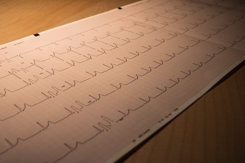
Electrocardiogram (ECG) — The Gold Standard
The diagnosis of Atrial Fibrillation (AFib) requires electrocardiographic confirmation. The diagnostic criteria for Atrial Fibrillation (AFib) on a 12-lead ECG are:
- Absence of distinct P waves — replaced by irregular fibrillatory (f) waves of varying morphology, amplitude, and frequency — best seen in lead V1 and the inferior leads
- Irregularly irregular R-R intervals — no consistent, repeating cycle length between QRS complexes
- Narrow QRS complexes — typically less than 120 milliseconds — unless aberrant conduction or a pre-existing bundle branch block is present
A single ECG tracing of at least 30 seconds showing the above criteria — or a 12-lead ECG interpreted by a physician — is required to establish the diagnosis of AF.
Ambulatory Electrocardiographic Monitoring
- 24-hour Holter Monitor — continuous ECG recording over 24-48 hours. Useful for frequent paroxysmal symptoms but may miss less frequent episodes.
- Extended Holter Monitoring (7- 14 days) significantly increases the diagnostic yield for paroxysmal AFib.
- Event Recorders — patient-activated devices that record ECG when symptoms occur.
- Implantable Loop Recorder (ILR) — a small subcutaneous device providing continuous ECG monitoring for up to 3 years. The gold standard for detecting very infrequent paroxysmal Atrial Fibrillation — particularly after cryptogenic stroke.
- Wearable Technology — consumer smartwatches with photoplethysmography (PPG) technology have demonstrated reasonable accuracy for Atrial Fibrillation detection, though all positive findings require confirmation with a physician-interpreted ECG.
Blood Tests
- Full Blood Count — to identify anaemia as a precipitant and assess for infection or inflammation.
- Thyroid Function Tests (TSH, Free T4) — mandatory in all new AF presentations to exclude hyperthyroidism as a reversible cause.
- Cardiac Biomarkers (Troponin, BNP/NT-proBNP) — elevated troponin suggests acute myocardial injury; elevated natriuretic peptides indicate elevated ventricular filling pressures and concurrent heart failure.
- Renal Function and Electrolytes — essential for identifying Chronic Kidney Disease and for guiding anticoagulation therapy. Electrolyte disturbances — hypokalaemia, hypomagnesaemia, hypocalcaemia — are important Atrial Fibrillation precipitants.
For a detailed clinical explanation of the various types of blood tests, please read our blood test article.
Echocardiography
- Transthoracic echocardiography (TTE) is recommended in all patients with newly diagnosed Atrial Fibrillation to assess ventricular size and function, identify structural heart disease, measure left atrial size, assess for LVH, and estimate pulmonary artery pressure.
- Transoesophageal Echocardiography (TOE) is the gold standard for imaging the left atrial appendage to exclude LAA thrombus before cardioversion or catheter ablation.
Stroke Risk Assessment — CHA2DS2-VASc Score
The CHA2DS2-VASc score is the internationally validated tool for quantifying stroke risk in AFib:
Congestive Heart Failure — 1 point, Hypertension — 1 point, Age: 75 years or above — 2 points, Diabetes Mellitus — 1 point, Stroke / Transient Ischemic Attack / Thromboembolism (prior) — 2 points, Vascular Disease (Coronary Artery Disease, Peripheral Artery Disease, Aortic Plaque) — 1 point, Age: 65-74 years — 1 point, Sex: Category Female — 1 point, Maximum Score — 9 points.
A CHA2DS2-VASc score of 2 or above in men or 3 or above in women indicates sufficient stroke risk to warrant oral anticoagulation therapy.
Bleeding Risk Assessment — HAS-BLED Score
The HAS-BLED score identifies modifiable bleeding risk factors before initiating anticoagulation. A score of 3 or above indicates high bleeding risk — importantly, a high HAS-BLED score should prompt correction of modifiable bleeding risk factors rather than automatic withholding of anticoagulation.
Treatment of Atrial Fibrillation

The treatment of Atrial Fibrillation is structured around four interconnected pillars — anticoagulation for stroke prevention, rate control, rhythm control, and upstream risk factor modification.
Pillar 1 — Anticoagulation for Stroke Prevention
Stroke prevention is the single most important therapeutic priority in Atrial Fibrillation management.
Direct Oral Anticoagulants
DOACs are the anticoagulants of choice for stroke prevention in non-valvular AFib — preferred over warfarin due to a superior safety profile, predictable pharmacokinetics, fixed dosing without routine monitoring, and a more rapid onset of action. Four DOACs are approved:
- Apixaban (Eliquis) — factor Xa inhibitor; twice a day dosing; superior to warfarin for both stroke prevention and major bleeding reduction.
- Rivaroxaban (Xarelto) — factor Xa inhibitor; once a day dosing; non-inferior to warfarin for stroke prevention with reduced intracranial haemorrhage.
- Dabigatran (Pradaxa) — direct thrombin inhibitor; twice a day dosing; reversible with idarucizumab.
- Edoxaban (Lixiana) — factor Xa inhibitor; once a day dosing.
All four DOACs significantly reduce the risk of intracranial haemorrhage compared to warfarin.
Warfarin
Warfarin remains the anticoagulant of choice in Atrial Fibrillation associated with rheumatic mitral stenosis or mechanical heart valves — where DOACs have not demonstrated equivalent efficacy. Requires regular Prothrombin Time Test (PT/INR) with a target of 2.0-3.0.
Left Atrial Appendage Occlusion (LAAO)
For patients with Atrial Fibrillation and high stroke risk who cannot tolerate long-term anticoagulation, percutaneous transcatheter occlusion of the left atrial appendage — using devices such as the Watchman — offers a non-pharmacological alternative for stroke risk reduction.
Pillar 2 — Rate Control
Rate control aims to reduce ventricular rate to prevent haemodynamic consequences of tachycardia. Current guidelines recommend a resting ventricular rate target below 110 beats per minute as an initial lenient target.
Beta-Blockers
Preferred first-line rate-control agents for most patients. Bisoprolol, metoprolol, and carvedilol are the most commonly used.
Non-Dihydropyridine Calcium Channel Blockers
Verapamil and diltiazem slow AV nodal conduction effectively. Contraindicated in heart failure with reduced ejection fraction.
Digoxin
Effective at rest but less effective during exercise. Used as an adjunct when rate control remains inadequate with other agents.
AV Node Ablation and Permanent Pacing
In patients with permanent Atrial Fibrillation refractory to pharmacological rate control, radiofrequency ablation of the AV node followed by permanent pacemaker implantation provides definitive rate control.
Pillar 3 — Rhythm Control
Rhythm control aims to restore and maintain sinus rhythm. The landmark EAST-AFNET 4 trial supports early rhythm control in symptomatic Atrial Fibrillation patients, demonstrating significant reductions in cardiovascular outcomes.
Pharmacological Cardioversion
Flecainide and Propafenone — class IC antiarrhythmics effective for cardioversion of recent-onset AFib in patients without structural heart disease. Flecainide can be used as a pill-in-the-pocket strategy for self-termination of infrequent paroxysmal AFib episodes.
Amiodarone — the most effective antiarrhythmic for cardioversion and rhythm maintenance, particularly in structural heart disease. Its use is limited by significant toxicity — pulmonary toxicity, thyroid dysfunction, hepatotoxicity, photosensitivity, corneal microdeposits, and peripheral neuropathy — requiring regular organ function monitoring.
Electrical Cardioversion (DC Cardioversion — DCCV)
Synchronised direct current electrical shock delivered under brief general anaesthesia — the most effective method of restoring sinus rhythm acutely. Restores sinus rhythm in approximately 90% of cases.
Important anticoagulation consideration: cardioversion of Atrial Fibrillation present for more than 48 hours requires either adequate anticoagulation for at least 3 weeks prior or a TOE to exclude LAA thrombus. Anticoagulation must continue for a minimum of 4 weeks after cardioversion due to atrial stunning.
Antiarrhythmic Drug Therapy for Rhythm Maintenance
Flecainide / Propafenone — first-line for patients without structural heart disease. Sotalol — class III antiarrhythmic; requires QT interval monitoring due to proarrhythmic risk. Dronedarone — non-iodinated amiodarone derivative; contraindicated in heart failure and permanent AFib. Amiodarone — most effective for rhythm maintenance in structural heart disease.
Catheter Ablation — Pulmonary Vein Isolation (PVI)
Catheter ablation has emerged as the most effective rhythm control strategy — superior to antiarrhythmic drug therapy for reducing Atrial Fibrillation recurrence and improving quality of life. The procedure involves advancing catheters through the femoral vein, crossing the interatrial septum via transseptal puncture into the left atrium, and delivering radiofrequency energy or cryotherapy to electrically isolate the pulmonary veins. Success rates are approximately 70-80% freedom from AFib at one year for paroxysmal AFib following a single procedure.
Surgical Ablation — The Cox-Maze Procedure
The Cox-Maze procedure creates a series of lesions in both atria, interrupting all potential macro-reentrant circuits. Long-term freedom from AF exceeds 90% in many series. Most commonly performed concomitantly with cardiac surgery for other indications.
Pillar 4 — Upstream Risk Factor Modification
Weight Loss — aggressive weight management reduces Atrial Fibrillation burden, symptom severity, and ablation recurrence rates. The LEGACY trial demonstrated that sustained weight loss of more than 10% significantly reduced AFib recurrence.
Blood Pressure Control — rigorous hypertension management reduces atrial remodelling and AFib recurrence.
Alcohol Reduction or Cessation — significantly reduces Atrial Fibrillation burden and recurrence.
Treatment of Obstructive Sleep Apnoea — Continuous Positive Airway Pressure therapy reduces Atrial Fibrillation recurrence rates and improves ablation outcomes.
Regular Moderate Exercise — moderate aerobic exercise is protective. The relationship between exercise intensity and Atrial Fibrillation is U-shaped — extreme endurance exercise may increase AFib risk.
Prognosis
Stroke — the most feared complication. Without anticoagulation, the annual stroke risk in Atrial Fibrillation patients with a CHA2DS2-VASc score of 2 or above is approximately 2-4% per year. Effective anticoagulation with DOACs reduces this risk by approximately 65-70%. AF-related strokes are typically large-territory, severely disabling, and carry a 30-day mortality of approximately 25%.
Heart Failure — Atrial Fibrillation increases the risk of heart failure hospitalization twofold. Successful rhythm control — particularly through catheter ablation — improves ventricular function and reduces hospitalisation rates.
Mortality — AFib is associated with approximately twofold increased all-cause mortality compared to age-matched individuals in sinus rhythm.
Quality of Life — symptomatic AFib significantly impairs quality of life. Catheter ablation consistently demonstrates superior improvements in quality of life compared to rate control alone.
Cognitive Decline and Dementia — Atrial Fibrillation is independently associated with increased risk of cognitive decline and dementia — even in the absence of clinically apparent stroke — through mechanisms including cerebral microembolism and cerebral hypoperfusion.
With optimal management — effective anticoagulation, adequate rate or rhythm control, and rigorous upstream risk factor modification — the prognosis of AFib has improved substantially over the past two decades.
Living With Atrial Fibrillation
Receiving a diagnosis of atrial fibrillation can be alarming — particularly when you understand the stroke risk it carries. But Atrial Fibrillation is one of the most actively managed cardiac conditions in medicine, with a growing and highly effective therapeutic armamentarium. The vast majority of people with well-treated AFib live full, active, and meaningful lives.
Take your anticoagulant therapy every single day without exception. If your CHA2DS2-VASc score warrants anticoagulation, this is the most important medication you take. The days you skip it are the days your stroke risk is unprotected. Modern DOACs are taken once or twice daily, are generally well tolerated, and do not require blood monitoring.
Know your triggers. Many patients with paroxysmal Atrial Fibrillation identify personal triggers — alcohol, caffeine, sleep deprivation, dehydration, intense stress, or illness. Keeping a symptom diary in the early months after diagnosis can identify your personal trigger pattern and reduce episode frequency through targeted lifestyle adjustments.
Monitor your heart rate and rhythm. A validated smartwatch or a personal ECG device allows you to monitor your own heart rhythm at home, detect AFib episodes, and share ECG recordings with your medical team.
Manage your lifestyle aggressively. The evidence that weight loss, alcohol reduction, blood pressure control, treatment of sleep apnoea, and regular moderate exercise reduce AFib burden is compelling and growing. Lifestyle modification is a core therapeutic intervention in AFib — not supplementary to medical treatment.
Understand that Atrial Fibrillation management is dynamic. AFib is a progressive condition in many patients — paroxysmal AFib frequently evolves to persistent AFib over years. Your treatment plan will evolve accordingly — from antiarrhythmic drugs to cardioversion to catheter ablation. This progression does not represent failure — it reflects the natural history of the condition and the availability of increasingly effective treatments at each stage.
Address the psychological impact. Living with an unpredictable arrhythmia generates significant anxiety in many patients. AF-related anxiety is real, clinically significant, and independently worsens outcomes. Seeking psychological support — through cardiac rehabilitation, counselling, or peer support groups — is a legitimate and important part of AF management.
Never stop anticoagulation without medical advice. Even if you feel well, even if you have been in sinus rhythm for months after cardioversion or ablation — never stop your anticoagulant without explicit guidance from your cardiologist. Silent, asymptomatic AF may be occurring without your awareness, maintaining your stroke risk at the same level as symptomatic AF.
When to Contact Emergency Services

Call emergency services immediately if you experience:
- Sudden onset of facial drooping, arm weakness, leg weakness, or speech difficulty — signs of stroke
- Sudden loss of vision in one or both eyes
- Sudden severe headache — the worst headache of your life
- Loss of consciousness or collapse
- Severe chest pain associated with breathlessness, sweating, or radiation to the arm or jaw
- Severe breathlessness at rest — possible acute pulmonary oedema from rapid Atrial Fibrillation
- Very rapid heart rate above 150 beats per minute associated with breathlessness, chest pain, presyncope, or hypotension
- Signs of major bleeding on anticoagulation — coughing up blood, vomiting blood, blood in urine or stools, or uncontrolled external bleeding
Contact your doctor or cardiologist urgently (same day) if you notice:
- A new or significantly more frequent or prolonged Atrial Fibrillation episode than your usual pattern
- Atrial Fibrillation that fails to terminate spontaneously within your usual timeframe
- New or worsening breathlessness, ankle swelling, or fatigue
- Palpitations associated with presyncope or near-fainting
- Signs of non-life-threatening bleeding on anticoagulation — unusual bruising, prolonged bleeding from minor cuts
- Missed doses of anticoagulant — particularly multiple consecutive missed doses
- Any new medication prescribed by another doctor — many common medications interact with anticoagulants
What to Ask Your Doctor About Atrial Fibrillation
About Your Diagnosis:
- What type of Atrial Fibrillation do I have — paroxysmal, persistent, or permanent — and what does this mean for my treatment options?
- What is my CHA2DS2-VASc score, and does my stroke risk warrant anticoagulation?
- Has an echocardiogram been performed to assess my heart structure and function?
- Has an underlying or reversible cause of my AFib been identified and addressed?
About Stroke Prevention:
- Which anticoagulant is most appropriate for me and at what dose?
- How does my kidney function affect my anticoagulant choice and dose?
- What should I do if I miss a dose of my anticoagulant?
- Are there any medications, foods, or supplements I should avoid while taking my anticoagulant?
About Rate and Rhythm Control:
- Is a rate control or rhythm control strategy more appropriate for me?
- Am I a candidate for cardioversion, and if so, when should it be performed?
- Am I a candidate for catheter ablation, and what success rate would you expect in my case?
- What antiarrhythmic medication is most appropriate for me given my heart structure and other conditions?
About Lifestyle:
- Which specific lifestyle modifications are most likely to reduce my Atrial Fibrillation burden?
- Is there evidence that my weight, alcohol intake, or sleep apnoea is contributing to my AFib?
- What level of physical activity is safe and beneficial for me?
About Monitoring and Follow-Up:
- How frequently should I be reviewed, and what investigations are needed at follow-up?
- Should I monitor my heart rhythm at home, and if so, what device do you recommend?
- What changes in my symptoms should prompt me to contact you urgently?
- At what point would you consider escalating my treatment from antiarrhythmic drugs to catheter ablation?
Sources
- Hindricks, G., et al. (2020). 2020 ESC Guidelines for the diagnosis and management of atrial fibrillation. European Heart Journal, 42(5), 373-498. https://doi.org/10.1093/eurheartj/ehaa612
- January, C.T., et al. (2019). 2019 AHA/ACC/HRS Focused Update of the 2014 Guideline for Management of Patients with Atrial Fibrillation. Journal of the American College of Cardiology, 74(1), 104-132. https://doi.org/10.1016/j.jacc.2019.01.011
- Kirchhof, P., et al. (2020). Early Rhythm-Control Therapy in Patients with Atrial Fibrillation (EAST-AFNET 4). New England Journal of Medicine, 383(14), 1305-1316. https://doi.org/10.1056/NEJMoa2019422
- Haissaguerre, M., et al. (1998). Spontaneous Initiation of Atrial Fibrillation by Ectopic Beats Originating in the Pulmonary Veins. New England Journal of Medicine, 339(10), 659-666. https://doi.org/10.1056/NEJM199809033391003
- Packer, D.L., et al. (2019). Catheter Ablation versus Antiarrhythmic Drug Therapy for Atrial Fibrillation (CABANA Trial). JAMA, 321(13), 1261-1274. https://doi.org/10.1001/jama.2019.0693
- Marrouche, N.F., et al. (2018). Catheter Ablation for Atrial Fibrillation with Heart Failure (CASTLE-AF). New England Journal of Medicine, 378(5), 417-427. https://doi.org/10.1056/NEJMoa1707855
- Lau, D.H., et al. (2017). Modifiable risk factors and atrial fibrillation. Circulation, 136(6), 583-596. https://doi.org/10.1161/CIRCULATIONAHA.116.023163
- Wolf, P.A., Abbott, R.D., and Kannel, W.B. (1991). Atrial fibrillation as an independent risk factor for stroke: the Framingham Study. Stroke, 22(8), 983-988. https://doi.org/10.1161/01.STR.22.8.983
- Granger, C.B., et al. (2011). Apixaban versus Warfarin in Patients with Atrial Fibrillation (ARISTOTLE Trial). New England Journal of Medicine, 365(11), 981-992. https://doi.org/10.1056/NEJMoa1107039
- Pisters, R., et al. (2010). A Novel User-Friendly Score (HAS-BLED) to Assess One-Year Risk of Major Bleeding in Atrial Fibrillation. Chest, 138(5), 1093-1100. https://doi.org/10.1378/chest.10-0134
- Linz, D., et al. (2018). Associations of obstructive sleep apnea with atrial fibrillation and continuous positive airway pressure treatment. JAMA Cardiology, 3(6), 532-540. https://doi.org/10.1001/jamacardio.2018.0095
- Abed, H.S., et al. (2013). Effect of weight reduction and cardiometabolic risk factor management on symptom burden and severity in patients with atrial fibrillation (LEGACY Trial). JAMA, 310(19), 2050-2060. https://doi.org/10.1001/jama.2013.280521
- Kasper, D.L., et al. (2018). Harrison’s Principles of Internal Medicine (20th ed.). McGraw-Hill Education.
Featured Image: Luan Rezende / Pexels
Copyright UncutMed. Medical knowledge truthful, precise, unfiltered. This article was last reviewed on September 3, 2026. Always consult your healthcare provider for personal medical advice.