© UncutMed — Medical knowledge, precise, truthful, unfiltered. This article was last reviewed on 10 August 2026. Always consult your healthcare professional for personal medical advice.
⚕️ Medical Disclaimer: This article is for informational and educational purposes only. It does not constitute medical advice, diagnosis, or treatment. Always consult a qualified healthcare professional regarding any medical condition or treatment plan.
Definition – What is Coronary Artery Disease?

The heart is a muscle that beats approximately 100,000 times each day. Just like every other muscle, it needs oxygen to keep contracting, fulfilling its vital role in maintaining blood circulation throughout the body so that every tissue and organ has a constant oxygenated blood supply. Three major blood vessels arising from the aortic sinuses supply the heart with oxygen-rich blood themselves.
Coronary Artery Disease — commonly abbreviated as CAD — is a condition in which these coronary arteries become narrowed or blocked, restricting the flow of blood to the heart muscle. The primary culprit is a process called atherosclerosis: the gradual buildup of a fatty, cholesterol-rich substance known as plaque inside the walls of the arteries. Over time, this plaque hardens, narrows the arterial channel, and makes the vessel walls stiffer and less flexible.
The result is a heart muscle that is chronically — or suddenly — starved of the oxygen it needs to function. Coronary Artery Disease is the most common form of heart disease worldwide and remains the single leading cause of death globally. Understanding it is not just medically important — it is potentially life-saving.
The Two Forms of Coronary Artery Disease
CAD does not present in a single and definitive way. It exists on a spectrum, broadly divided into two major clinical forms based on how the disease manifests and how quickly it develops.
Chronic Coronary Syndrome (Stable CAD)
Chronic Coronary Syndrome — previously known as stable angina or stable ischemic heart disease — represents the slow, progressive form of CAD. In this form, plaque builds up gradually inside the coronary arteries over many years, slowly narrowing the vessel and reducing blood flow to the heart muscle.
In stable Coronary Artery Disease, the narrowing is significant but consistent. The heart receives ample blood flow at rest, but during periods of increased demand — physical exercise, emotional stress, cold weather — the narrowed artery cannot deliver enough oxygen-rich blood to meet that demand. This mismatch between oxygen supply and demand produces the characteristic symptoms of stable angina: chest discomfort that is predictable, reproducible, and relieved by rest or medication.
Chronic Coronary Syndrome is manageable with medication and lifestyle modification, but it represents an important warning sign that the coronary arteries are significantly narrowed and diseased, so that the risk of a more acute event is elevated.
Acute Coronary Syndrome (ACS)
Acute Coronary Syndrome represents the dangerous, rapidly evolving end of the CAD spectrum. It occurs when a plaque inside a coronary artery suddenly ruptures or erodes. The body reacts to this rupture as it would to any injury — by forming a blood clot at the site. If this clot partially or completely blocks the artery, blood flow to the heart muscle is abruptly cut off.
Acute Coronary Syndrome (ACS) is an umbrella term that encompasses three distinct but closely related conditions, classified based on the severity of the blockage and the degree of heart muscle damage:
- Unstable Angina: The clot partially blocks the artery, causing chest pain that occurs at rest or with minimal physical exertion — unpredictable and more severe than stable angina. No permanent heart muscle damage occurs, but it is a medical emergency requiring immediate attention.
- Non-ST Elevation Myocardial Infarction (NSTEMI): The artery is significantly but NOT completely blocked. A NSTEMI happens when a blood clot forms over plaque in one of the coronary arteries, causing a partial blockage. An NSTEMI involves partial or transiently collateralized occlusion causing inner-wall (subendocardial) injury. Blood flow is severely reduced, and heart muscle begins to sustain damage, detectable through elevated cardiac biomarkers in the blood (particularly troponin). This condition gets its name because it doesn’t cause a specific electrical pattern called ST-segment elevation on an electrocardiogram (ECG/EKG) test. This phenomenon in the ECG differentiates a non-STEMI from a STEMI (more information below).
- ST Elevation Myocardial Infarction (STEMI): The artery is completely blocked. STEMI involves a total blockage across the full thickness of the heart muscle wall. STEMI results in injury through the entire wall of the heart muscle (transmural ischemia), creating an electrical injury current that shifts the ECG baseline upward. Blood flow to a section of the heart muscle is entirely cut off, causing rapid and extensive heart muscle death. This is what most people refer to as a “heart attack” and represents the most severe and time-critical form of ACS. In a STEMI, every minute without treatment results in further irreversible muscle loss — a principle captured in the clinical phrase: “time is muscle.”
Symptoms and Signs of Coronary Artery Disease
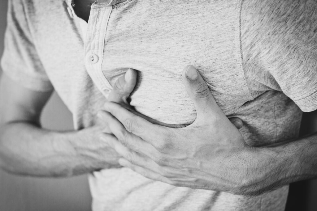
The symptoms of Coronary Artery Disease vary considerably depending on whether the presentation is chronic or acute. It is also critically important to note that Coronary Artery Disease can be entirely silent — particularly in its early stages and in certain populations such as diabetic patients and women — making awareness of the full symptom spectrum essential.
Symptoms of Chronic Coronary Artery Disease (Stable Angina)
- Chest pain or discomfort (Angina): The most typical symptom of stable Coronary Artery Disease. Most of the time, it is described as a pressure, tightness, squeezing, or heaviness in the centre of the chest. Many patients express their discomfort using Levine’s sign. This is a physical gesture where a patient places a fist, often against the breastbone (sternum), to visually describe a crushing, squeezing, or pressing discomfort associated with cardiac events like a heart attack or angina. The feeling of chest pain/discomfort is provoked by physical exercise or stress and relieved within minutes by rest or sublingual nitrates. It does not typically change with breathing or body position.
- Radiation of discomfort: The chest discomfort of angina frequently radiates to the left arm, jaw, neck, shoulder, or back — reflecting the shared nerve pathways between the heart and these regions. This means that the pain/discomfort has the chest as its main location, but then it is extended to body areas that share the same nerve distribution.
- Shortness of breath (Dyspnoea): Occurs because a poorly perfused heart muscle pumps less efficiently, causing fluid to back up into the lungs, creating the feeling of having difficulty breathing. May occur alongside chest pain or in isolation — particularly in elderly patients and women.
- Fatigue: Chronic reduction in cardiac output leads to persistent fatigue and reduced exercise tolerance. Many patients unconsciously reduce their activity levels to avoid triggering symptoms, masking the true severity of their disease.
- Dizziness or lightheadedness: Reduced cardiac output can temporarily reduce blood flow to the brain, causing dizziness — particularly during physical exercise.
Symptoms of Acute Coronary Syndrome
- Severe, prolonged chest pain: Unlike stable angina, the chest pain of Acute Coronary Syndrome is more intense, lasts longer than 20 minutes, occurs at rest, and is not relieved by nitrates or rest. It may be described as crushing or a sensation of impending doom.
- Diaphoresis (profuse sweating): A sudden, cold sweat accompanying chest pain is a classic and important warning sign of Acute Coronary Syndrome. This mechanism is driven by the activation of the sympathetic nervous system in response to cardiac stress.
- Nausea and vomiting: Common in inferior myocardial infarctions due to stimulation of vagal nerve pathways. The Vagal Reflex is triggered when damaged heart tissue and ischemic metabolites (such as lactic acid) stimulate cardiac sensory receptors. This activates the vagus nerve, sending distress signals that provoke a parasympathetic gastrointestinal response. The sudden cardiac event forces the body to release high levels of catecholamines (stress hormones), which disrupt normal stomach and gut motility.
- Palpitations: Awareness of rapid, irregular, or forceful heartbeats — reflecting arrhythmias that commonly accompany acute ischaemia in the coronary arteries.
- Syncope (loss of consciousness): Can occur due to a dangerous arrhythmia or severely reduced cardiac output during an acute event.
Atypical Presentations — Particularly Important
A significant proportion of patients — especially women, elderly individuals, and those with diabetes — present with atypical symptoms that do not include classic chest pain. These may include:
- Unexplained jaw or arm pain
- Nausea without chest pain
- Unusual fatigue or weakness
- Shortness of breath as the only symptom
- Epigastric (upper abdominal) discomfort mistaken for indigestion
These atypical presentations are a major reason why Coronary Artery Disease is underdiagnosed and undertreated in certain populations. Any unexplained symptom in a person with known risk factors for Coronary Artery Disease warrants prompt medical evaluation.
Causation – What Causes Coronary Artery Disease?
The underlying cause of CAD in the vast majority of cases is atherosclerosis — a complex, chronic inflammatory process that begins in the walls of the arteries and progresses over decades.
The process unfolds in several stages:
- Endothelial Injury: The inner lining of the coronary artery — the endothelium — is exposed to damaging forces, including high blood pressure, high blood sugar, smoking toxins, and oxidised LDL cholesterol. This causes microscopic injury to the endothelial surface, making it permeable and dysfunctional.
- LDL Infiltration and Oxidation: LDL cholesterol particles penetrate the damaged endothelium and become trapped within the artery wall. Once inside, they undergo oxidation — a chemical change that makes them highly inflammatory and toxic to surrounding tissue.
- Inflammatory Response: The immune system recognises oxidised LDL as an unsafe, foreign substance. Monocytes from the bloodstream migrate into the artery wall and transform into macrophages, which engulf the oxidised LDL. These engorged macrophages become “foam cells” — the earliest building block of atherosclerotic plaque.
- Plaque Formation: Over time, foam cells accumulate, smooth muscle cells migrate into the area, and fibrous tissue forms around the growing lipid core. This creates an atherosclerotic plaque — a raised, fatty deposit within the artery wall that progressively narrows the vessel lumen.
- Plaque Rupture and Thrombosis: Plaques with a thin fibrous cap and a large lipid core are particularly vulnerable to rupture. The higher the fibrous element of the plaque, the lower the risk of rupture is. The higher the lipid element of the plaque, the higher the risk of rupture is. When the fibrous cap of the plaque tears, the lipid core is exposed to blood, triggering immediate clot formation. It is this acute thrombosis — not the plaque itself — that causes the sudden complete blockage of the coronary arteries responsible for a heart attack.
Risk Factors for Coronary Artery Disease

Coronary Artery Disease is a multifactorial disease — meaning it results from the interplay of multiple risk factors rather than a single cause. Risk factors are divided into modifiable (those you can change) and non-modifiable (those you cannot).
Non-Modifiable Risk Factors
- Age: The risk of Coronary Artery Disease increases substantially with age. Atherosclerosis is a lifelong process, and its clinical consequences typically emerge after decades of progression. Men over 45 and women over 55 are at significantly increased risk.
- Sex: Men develop Coronary Artery Disease earlier than women. However, after menopause, women’s risk rises sharply — by the age of 65, the risk becomes approximately equal between the two sexes. Oestrogen is believed to have a protective effect on the cardiovascular system before menopause.
- Family History and Genetics: A first-degree relative (parent or sibling) who developed Coronary Artery Disease before the age of 55 (men) or 65 (women) significantly elevates personal risk. Several genetic variants have been identified that influence cholesterol metabolism, blood pressure regulation, and inflammatory responses.
- Ethnicity: Certain ethnic groups carry higher baseline risk. South Asian populations, for example, have a disproportionately high incidence of Coronary Artery Disease at younger ages compared to other ethnic groups.
- Early menopause (before age 40): The timing of menopause is largely biological and cannot be controlled. As stated above, this is linked to the potential protective effect of oestrogen in the cardiovascular system before menopause.
- History of gestational diabetes, eclampsia, or preeclampsia: These are past events that already occurred and cannot be undone, though their risk can be managed going forward.
- Endometriosis: A chronic condition that cannot be fully reversed.
Modifiable Risk Factors
- Hypertension (High Blood Pressure): Chronically elevated blood pressure exerts mechanical stress on the arterial endothelium, accelerating the process of endothelial damage and atherosclerosis. Hypertension is one of the single most powerful modifiable risk factors for Coronary Artery Disease.
- Dyslipidaemia (Abnormal Cholesterol): Elevated LDL cholesterol and low HDL cholesterol are strongly associated with accelerated atherosclerosis progression. Elevated triglycerides also contribute to cardiovascular risk.
- Diabetes Mellitus: Chronically elevated blood glucose damages the endothelium directly, promotes inflammation, and accelerates atherosclerosis. Diabetic patients have a two to four times higher risk of developing CAD compared to non-diabetic individuals.
- Smoking: Tobacco smoke contains thousands of toxic chemicals that damage the endothelium, reduce HDL cholesterol, increase LDL oxidation, promote blood clotting, and cause coronary artery spasm. Smokers have a two to four times higher risk of Coronary Artery Disease than non-smokers. Importantly, this risk begins to fall within months of cessation.
- Obesity: Excess body weight — particularly central (abdominal) obesity — is associated with hypertension, dyslipidaemia, insulin resistance, and systemic inflammation, all of which independently accelerate Coronary Artery Disease.
- Physical Inactivity: A sedentary lifestyle is independently associated with increased cardiovascular risk. Regular physical activity improves lipid profiles, reduces blood pressure, aids weight control, and improves insulin sensitivity.
- Unhealthy Diet: A diet high in saturated fats, trans fats, refined carbohydrates, and excess sodium promotes dyslipidaemia, hypertension, obesity, and systemic inflammation — all key drivers of atherosclerosis.
- Chronic Stress: Psychological stress activates the sympathetic nervous system, raises blood pressure, promotes inflammation, and is associated with unhealthy coping behaviours such as smoking and overeating. Chronic stress is an increasingly recognised independent risk factor for Coronary Artery Disease.
- Poor sleep: Not sleeping enough, as well as sleep disorders such as sleep apnea. Obstructive sleep apnea — the most common sleep disorder — causes repeated episodes of oxygen deprivation during sleep as the airway intermittently collapses. Each apnoeic episode triggers a surge in sympathetic nervous system activity, causing spikes in heart rate and blood pressure throughout the night. Over time, this chronic nocturnal cardiovascular stress causes sustained hypertension, promotes systemic inflammation, increases platelet aggregability, and accelerates atherosclerosis.
- Excessive Alcohol Consumption: While moderate alcohol consumption has been debated as potentially cardioprotective, excessive intake raises blood pressure, promotes arrhythmias, and contributes to obesity and dyslipidaemia.
- Autoimmune diseases (lupus, rheumatoid arthritis): Chronic systemic inflammation is the key mechanism linking autoimmune diseases to cardiovascular risk. These conditions cannot be fully cured but can be managed with medication, reducing their cardiovascular impact.
- HIV/AIDS: Even when well controlled with modern antiretroviral therapy, it creates a state of chronic immune activation and systemic inflammation that accelerates atherosclerosis. The virus itself promotes endothelial dysfunction and alters lipid metabolism, raising LDL and triglycerides while lowering HDL.
- Hormonal Birth Control: Combined oral contraceptives — containing both oestrogen and progestogen — raise cardiovascular risk primarily through two mechanisms. First, oestrogen promotes a prothrombotic state by increasing clotting factors and reducing natural anticoagulants, raising the risk of arterial and venous thrombosis. Second, combined contraceptives can raise blood pressure and adversely alter lipid profiles in susceptible individuals.
- Anemia: Anemia reduces the oxygen-carrying capacity of the blood. To compensate, the heart increases its output — beating faster and harder to deliver adequate oxygen to tissues. This chronic increase in cardiac workload accelerates wear on the coronary arteries, raises myocardial oxygen demand, and can trigger angina in patients with pre-existing Coronary Artery Disease. Severe anemia can even precipitate a myocardial infarction in the absence of significant atherosclerosis — a phenomenon known as Type 2 MI.
- Chronic Kidney Disease (CKD): The kidneys and cardiovascular system are deeply interconnected — a relationship so well established that cardiologists and nephrologists refer to it as the cardiorenal syndrome. CKD contributes to cardiovascular risk through multiple simultaneous mechanisms. It causes fluid retention and hypertension, promotes dyslipidaemia, leads to anaemia, generates a chronic inflammatory state, and causes abnormal calcium and phosphate metabolism that directly calcifies arterial walls.
Complications of Coronary Artery Disease
When left untreated or inadequately managed, Coronary Artery Disease can lead to a range of serious and potentially life-threatening complications:
- Myocardial Infarction (Heart Attack): The most feared acute complication — complete blockage of a coronary artery leads to irreversible death of heart muscle tissue. The extent of damage depends on which artery is blocked and how quickly treatment is received.
- Heart Failure: Repeated episodes of ischaemia or a large myocardial infarction can permanently weaken the heart muscle, reducing its pumping capacity. The result is heart failure — a chronic condition in which the heart cannot meet the body’s demands for blood and oxygen.
- Arrhythmias: Ischaemic damage to the heart’s electrical conduction system can cause dangerous abnormal heart rhythms. Ventricular fibrillation — a disorganised, chaotic rhythm that stops effective pumping — is the most common cause of sudden cardiac death.
- Sudden Cardiac Death: Coronary Artery Disease is the underlying cause in the majority of cases of sudden cardiac death — defined as unexpected death from cardiac causes within one hour of symptom onset. It is most commonly caused by a lethal arrhythmia triggered by acute ischaemia.
- Cardiogenic Shock: A severe complication of massive myocardial infarction in which the heart muscle is so extensively damaged that it can no longer maintain adequate blood pressure and organ perfusion. Cardiogenic shock carries a very high mortality rate.
- Stroke: Coronary Artery Disease reflects widespread atherosclerosis affecting not just the coronary arteries but blood vessels throughout the body. Patients with CAD have a significantly elevated risk of ischaemic stroke due to atherosclerosis of the cerebral vessels or clots originating from a damaged heart.
Diagnostics and Tests – How is Coronary Artery Disease diagnosed

Diagnosing CAD involves a combination of clinical assessment, non-invasive testing, and in some cases, invasive procedures. The choice of investigations depends on the clinical presentation — whether the patient presents with stable symptoms or an acute event.
Clinical Assessment
- Medical History: The diagnostic process always begins with a thorough medical history. The physician will ask detailed questions about the nature, location, duration, and triggers of any chest discomfort, alongside a full assessment of cardiovascular risk factors, including smoking history, family history, blood pressure, cholesterol, and diabetes status.
- Physical Examination: A focused cardiovascular examination is performed, including measurement of blood pressure in both arms, heart rate, assessment of heart sounds for murmurs or added sounds, examination of the neck veins, and inspection of the extremities for signs of poor circulation or fluid retention.
Blood tests
- Cardiac Biomarkers: Troponin I and Troponin T are proteins released into the bloodstream when heart muscle cells are damaged. They are the cornerstone blood tests for diagnosing myocardial infarction. High-sensitivity troponin assays can detect even minor degrees of heart muscle injury within 1 to 3 hours of symptom onset.
- Lipid Panel: Assessment of total cholesterol, LDL, HDL, and triglycerides to evaluate atherogenic risk.
- Fasting Glucose and HbA1c: To screen for diabetes and prediabetes as contributing risk factors.
- Full Blood Count, Renal Function, and Thyroid Function: To identify contributing conditions such as anaemia, chronic kidney disease, or thyroid dysfunction.
Non-Invasive Cardiac Tests
- Electrocardiogram (ECG): A 12-lead ECG is one of the first and most important tests performed in any patient with suspected Coronary Artery Disease. It records the heart’s electrical activity and can identify characteristic changes associated with ischaemia, myocardial infarction, and arrhythmias. In a STEMI, the ECG shows the classic ST segment elevation that gives the condition its name.
- Exercise Stress Test (Exercise ECG): The patient walks on a treadmill or cycles on a stationary bike while connected to an ECG monitor. As heart rate and oxygen demand increase with exercise, areas of the heart receiving insufficient blood flow produce characteristic ECG changes — revealing CAD that may not be apparent at rest.
- Echocardiogram: An ultrasound of the heart that provides real-time images of the heart’s structure and function. It assesses the pumping function of the heart (ejection fraction), identifies areas of the heart muscle that are not contracting normally due to ischaemia or infarction, and evaluates the heart valves.
- Stress Echocardiogram: Combines an echocardiogram with physical or pharmacological stress (using medications such as dobutamine to simulate exercise). Wall motion abnormalities that appear under stress but not at rest indicate significant CAD.
- Myocardial Perfusion Imaging (MPI) — Nuclear Stress Test: A small amount of radioactive tracer is injected into the bloodstream. A special camera captures images of blood flow through the heart muscle both at rest and under stress, identifying areas of reduced perfusion that indicate significant coronary artery narrowing.
- CT Coronary Calcium Score: A non-contrast CT scan that detects and quantifies calcified plaque within the coronary arteries. The resulting score — the Agatston score — is a powerful predictor of cardiovascular risk, particularly useful in intermediate-risk patients where the decision to start preventive medication is uncertain.
- CT Coronary Angiography (CTCA): A contrast-enhanced CT scan that produces detailed three-dimensional images of the coronary arteries, allowing non-invasive visualisation of both calcified and non-calcified plaques and the degree of arterial narrowing. Increasingly used as a first-line investigation for stable chest pain in lower-risk patients.
Invasive Procedures
- Invasive Coronary Angiography (Cardiac Catheterisation): The gold standard for diagnosing and evaluating CAD. A catheter is threaded through an artery in the wrist or groin to the coronary arteries, and contrast dye is injected while X-ray imaging captures real-time pictures of blood flow through the coronary arteries. It precisely identifies the location and severity of blockages and is performed immediately before PCI when intervention is planned.
- Fractional Flow Reserve (FFR): Performed during invasive angiography, FFR uses a pressure wire passed across a narrowing in a coronary artery to measure the functional significance of that narrowing — determining whether it is severe enough to actually limit blood flow and warrant intervention. It prevents unnecessary stenting of lesions that look significant on angiography but are not functionally important.
- Intravascular Ultrasound (IVUS) and Optical Coherence Tomography (OCT): Advanced intracoronary imaging techniques performed during cardiac catheterisation that provide highly detailed cross-sectional images of the artery wall. They are used to characterise plaque composition, guide stent deployment, and assess stent results after PCI.
Treatment of Coronary Artery Disease

The treatment of CAD is divided into pharmaceutical (medication-based) and surgical/interventional approaches. In most patients, a combination of both is employed alongside lifestyle modification.
Pharmaceutical Treatment
- Antiplatelet Agents: Medications such as aspirin and clopidogrel reduce the ability of platelets to coagulate and form blood clots within narrowed coronary arteries. Aspirin is the cornerstone of CAD medical therapy and is recommended for virtually all patients with established CAD. Dual antiplatelet therapy (aspirin combined with a second agent such as clopidogrel, ticagrelor, or prasugrel) is standard following ACS or coronary stenting.
- Statins: Statins (e.g., atorvastatin, rosuvastatin) are among the most important medications in cardiovascular medicine. They reduce LDL cholesterol by inhibiting its production in the liver, slow down the progression of atherosclerotic plaques, stabilise vulnerable plaques, reducing rupture risk, and have anti-inflammatory effects on the vessel wall. All patients with established CAD should be on high-intensity statin therapy unless contraindicated.
- Beta-Blockers: Beta-blockers (e.g., bisoprolol, metoprolol, carvedilol) reduce heart rate and blood pressure, decreasing the heart’s oxygen demand. They are highly effective in managing stable angina, reducing the frequency and severity of episodes. Following a myocardial infarction, they significantly reduce the risk of death and recurrent events.
- Nitrates: Nitrates (e.g., sublingual glyceryl trinitrate, isosorbide mononitrate) cause dilation of the coronary arteries and peripheral blood vessels, reducing blood pressure, decreasing the heart’s workload, and relieving angina symptoms rapidly. Short-acting sublingual nitrates are used for immediate symptom relief during angina attacks. Long-acting nitrates are used for prevention.
- ACE Inhibitors and ARBs: Angiotensin-converting enzyme (ACE) inhibitors (e.g., ramipril, lisinopril) and angiotensin receptor blockers (ARBs) reduce blood pressure, decrease the workload on the heart, and have protective effects on the heart muscle following myocardial infarction. They are particularly important in patients with CAD who also have heart failure, hypertension, or diabetes.
- Calcium Channel Blockers: Calcium channel blockers (e.g., amlodipine, diltiazem) relax the smooth muscle of coronary and peripheral arteries, reducing blood pressure and relieving angina. They are particularly useful when beta-blockers are contraindicated or insufficient alone.
- Anticoagulants: In the acute setting of ACS, anticoagulants such as heparin or low-molecular-weight heparin are used to prevent further clot propagation. In patients with certain arrhythmias such as atrial fibrillation — common in CAD patients — oral anticoagulants such as warfarin or direct oral anticoagulants (DOACs) are used to prevent stroke.
- PCSK9 Inhibitors: A newer class of powerful cholesterol-lowering medications (e.g., evolocumab, alirocumab) used in patients who cannot achieve adequate LDL reduction with statins alone or who are statin-intolerant. They can reduce LDL cholesterol by up to 60% beyond what statins achieve.
Surgical and Interventional Treatment
- Percutaneous Coronary Intervention (PCI) — Coronary Angioplasty and Stenting: PCI is the most commonly performed interventional procedure for CAD. A thin flexible tube (catheter) is threaded through an artery in the wrist or groin up to the blocked coronary artery. A small balloon is inflated at the site of blockage to compress the plaque and widen the vessel — a process called angioplasty. A small metal mesh tube called a stent is then deployed to hold the artery open permanently. Modern drug-eluting stents are coated with medication that prevents the artery from re-narrowing (restenosis). In ACS — particularly STEMI — emergency PCI (primary PCI) is the gold standard treatment, ideally performed within 90 minutes of first medical contact.
- Coronary Artery Bypass Grafting (CABG): CABG is open-heart surgery in which blood vessels harvested from elsewhere in the body — typically the internal mammary artery from the chest wall or the saphenous vein from the leg — are used to create new routes (bypasses) around blocked coronary arteries. CABG is preferred over PCI in patients with multiple vessel disease, left main coronary artery disease, or in diabetic patients with extensive CAD where surgical revascularisation has been shown to provide superior long-term outcomes. Recovery from CABG is longer than PCI, but the durability of the bypass grafts — particularly arterial grafts — is excellent.
- Thrombolysis (Clot-Busting Therapy): In settings where primary PCI is not immediately available — such as remote areas — thrombolytic medications (e.g., alteplase, tenecteplase) can be administered intravenously to dissolve the clot causing a STEMI. While less effective than primary PCI, it is a critical life-saving option when catheterisation laboratory facilities are not accessible within the recommended timeframe.
- Implantable Cardioverter Defibrillator (ICD): In patients whose CAD has caused significant heart muscle damage with a high risk of life-threatening arrhythmias, an ICD may be implanted. This small device continuously monitors heart rhythm and delivers an electric shock to restore normal rhythm if a dangerous arrhythmia is detected — effectively acting as an internal defibrillator.
- Cardiac Rehabilitation: Following any major cardiac event or procedure, structured cardiac rehabilitation — combining supervised exercise, education, and psychological support — is an essential component of recovery. It significantly reduces the risk of future events, improves functional capacity, and enhances quality of life.
Prevention and Lowering Your Risk
Coronary Artery Disease is largely a preventable disease. The same risk factors that drive its development are modifiable — and the evidence for lifestyle intervention in reducing cardiovascular risk is among the strongest in all of medicine. Prevention and treatment in CAD are inseparable. The lifestyle modifications outlined in this section are not merely preventive measures for those yet to develop the disease — they are clinically proven therapeutic interventions for patients with established CAD. Dietary improvement, regular physical activity, smoking cessation, and weight management directly reduce angina symptoms, slow atherosclerosis progression, improve cardiac function, and reduce the risk of future events. In patients with early or mild CAD, aggressive lifestyle modification alone can halt and, in some cases, partially reverse disease progression. They should therefore be considered an active and essential component of every CAD management plan — not optional additions to medication.
- Quit Smoking: Smoking cessation is the single most impactful lifestyle change a person with CAD risk can make. Within one year of quitting, the excess risk of CAD is halved. Within 15 years, it approaches that of a lifelong non-smoker. Support through nicotine replacement therapy, varenicline, or bupropion significantly improves cessation success rates.
- Adopt a Heart-Healthy Diet: The Mediterranean diet — rich in vegetables, fruits, whole grains, legumes, fish, and olive oil, with limited red meat and processed foods — has the strongest evidence base for cardiovascular protection. Reducing saturated fat, trans fat, added sugars, and excess sodium directly improves lipid profiles and blood pressure.
- Exercise Regularly: Current guidelines recommend at least 150 minutes of moderate-intensity aerobic exercise per week — such as brisk walking, cycling, or swimming. Regular physical activity lowers blood pressure, raises HDL cholesterol, aids weight management, improves insulin sensitivity, and reduces systemic inflammation.
- Maintain a Healthy Weight: Achieving and maintaining a healthy body mass index (BMI of 18.5–24.9 kg/m²) and waist circumference (below 94cm in men and 80cm in women) significantly reduces cardiovascular risk by improving blood pressure, lipid profiles, and glucose metabolism. BMI can sometimes be misleading (such as in individuals with high muscle mass), but nonetheless it is a good indicator for body weight. Lipid allocation is a more useful index.
- Control Blood Pressure: Target blood pressure in most adults should be below 130 (systolic)/80 (diastolic) mmHg. This is achieved through lifestyle modification and medication when necessary. Regular monitoring — including home blood pressure measurement — is essential. Blood pressure must be measured at the same time every day and under certain conditions for the results to be consistent, truthful and comparable.
- Manage Cholesterol: Regular lipid panel (LDL, HDL, triglycerides) monitoring allows early identification and treatment of dyslipidaemia. Dietary modification is the first step, followed by statin therapy when indicated by cardiovascular risk assessment.
- Manage Diabetes: Tight glycaemic control in diabetic patients — with target HbA1c typically below 7% — significantly reduces the rate of cardiovascular complications. Newer diabetes medications, including GLP-1 receptor agonists (e.g. semaglutide) and SGLT-2 inhibitors (e.g. empagliflozin), have demonstrated direct cardiovascular protective effects beyond glucose lowering.
- Manage Stress: Incorporating stress-reduction strategies — mindfulness, adequate sleep, psychological support, and social connection into daily life has measurable benefits for cardiovascular health. Chronic psychological stress is a modifiable and frequently overlooked cardiovascular risk factor.
- Limit Alcohol: Current cardiovascular guidelines recommend limiting alcohol to no more than 14 units per week for both men and women, spread across several days, with alcohol-free days each week.
- Regular Medical Check-ups: Routine cardiovascular risk assessment — including blood pressure measurement, lipid panel, fasting glucose, and BMI — allows early identification of risk factors and timely intervention before CAD develops or progresses.
Living With Coronary Artery Disease — What to Expect
A diagnosis of Coronary Artery Disease is life-changing — but it is not a life sentence. With appropriate treatment, consistent lifestyle modification, and regular medical follow-up, the vast majority of people with CAD lead full, active, and meaningful lives.
It is a chronic condition requiring long-term management.
CAD does not disappear with treatment — atherosclerosis is a lifelong process. Medications will likely be taken indefinitely, and lifestyle habits maintained consistently. The goal of treatment is to slow progression, prevent complications, and maximise quality of life.
Symptoms can be well controlled.
Most patients with stable CAD achieve excellent symptom control with medication, allowing them to engage in normal daily activities, exercise, and work without significant limitation.
The risk of future events can be substantially reduced.
Adherence to medical therapy and lifestyle modification dramatically reduces the risk of heart attack, stroke, and cardiovascular death. The prognosis for a person with well-managed CAD who does not smoke, maintains a healthy lifestyle, and takes their medications consistently is significantly better than many people expect at diagnosis.
Emotional and psychological wellbeing matters.
A diagnosis of heart disease is associated with increased rates of anxiety and depression — both of which independently worsen cardiovascular outcomes. Addressing mental health as part of comprehensive CAD management is not optional — it is medically important. Cardiac rehabilitation programmes incorporate psychological support for this reason.
Regular follow-up is essential.
Routine monitoring of blood pressure, lipid levels, heart function, and medication tolerance allows your medical team to adjust treatment as your condition evolves. Do not skip follow-up appointments — they are a core part of managing this condition safely.
Know your warning signs.
This isn’t just important; it is vital. Every person with Coronary Artery Disease should know the symptoms of Acute Coronary Syndrome (look at the respective section of this article for more information) — particularly chest pain at rest, worsening angina, or new shortness of breath — and understand that these require immediate emergency medical attention. Time is muscle.
Living with Coronary Artery Disease demands awareness, consistency, and partnership with your healthcare team. It also demands self-compassion — managing a chronic condition is not easy, and small, sustained steps in the right direction are far more valuable than perfection.
Sources
- Virani, S.S., et al. (2021). Heart Disease and Stroke Statistics — 2021 Update. Circulation, 143(8), e254–e743. https://doi.org/10.1161/CIR.0000000000000950
- Knuuti, J., et al. (2019). 2019 ESC Guidelines for the diagnosis and management of chronic coronary syndromes. European Heart Journal, 41(3), 407–477. https://doi.org/10.1093/eurheartj/ehz425
- Collet, J.P., et al. (2020). 2020 ESC Guidelines for the management of acute coronary syndromes in patients presenting without persistent ST-segment elevation. European Heart Journal, 42(14), 1289–1367. https://doi.org/10.1093/eurheartj/ehaa575
- Ibanez, B., et al. (2017). 2017 ESC Guidelines for the management of acute myocardial infarction in patients presenting with ST-segment elevation. European Heart Journal, 39(2), 119–177. https://doi.org/10.1093/eurheartj/ehx393
- Grundy, S.M., et al. (2019). 2018 AHA/ACC Guideline on the Management of Blood Cholesterol. Journal of the American College of Cardiology, 73(24), e285–e350. https://doi.org/10.1016/j.jacc.2018.11.003
- Whelton, P.K., et al. (2018). 2017 ACC/AHA Guideline for the Prevention, Detection, Evaluation, and Management of High Blood Pressure in Adults. Journal of the American College of Cardiology, 71(19), e127–e248. https://doi.org/10.1016/j.jacc.2017.11.006
- Libby, P. (2021). The changing landscape of atherosclerosis. Nature, 592, 524–533. https://doi.org/10.1038/s41586-021-03392-8
- Estruch, R., et al. (2018). Primary Prevention of Cardiovascular Disease with a Mediterranean Diet Supplemented with Extra-Virgin Olive Oil or Nuts. New England Journal of Medicine, 378(25), e34. https://doi.org/10.1056/NEJMoa1800389
- Piepoli, M.F., et al. (2016). 2016 European Guidelines on cardiovascular disease prevention in clinical practice. European Heart Journal, 37(29), 2315–2381. https://doi.org/10.1093/eurheartj/ehw106
- Neumann, F.J., et al. (2018). 2018 ESC/EACTS Guidelines on myocardial revascularization. European Heart Journal, 40(2), 87–165. https://doi.org/10.1093/eurheartj/ehy394
- Kasper, D.L., et al. (2018). Harrison’s Principles of Internal Medicine (20th ed.). McGraw-Hill Education.
- World Heart Federation. (2023). Coronary Artery Disease. https://world-heart-federation.org/world-heart-day/facts-about-heart-disease/
© UncutMed — Medical knowledge, precise, truthful, unfiltered. This article was last reviewed on 10 August 2026. Always consult your healthcare professional for personal medical advice.