© UncutMed — Medical knowledge, precise, truthful, unfiltered. The current article was last modified on September 8, 2026. This article was clinically authored and reviewed under international evidence-based standards. Always consult your personal healthcare physician before modifying any medical therapy.
⚕️ Medical Disclaimer: This article is for informational and educational purposes only. It does not constitute medical advice, formal diagnosis, or treatment recommendations. Always consult a qualified healthcare professional regarding any metabolic condition or therapeutic plan.
1. Diabetes Mellitus Type 2 – Clinical Definition
Section Summary:
Type 2 Diabetes Mellitus is a progressive cardiometabolic disorder characterized by peripheral insulin resistance paired with progressive pancreatic beta-cell secretory failure. The resulting chronic hyperglycemia causes widespread endothelial injury, accelerated atherosclerosis, microvascular damage, and multi-organ dysfunction, forming the core metabolic driver of Cardiovascular-Kidney-Metabolic (CKM) syndrome.

Type 2 Diabetes Mellitus (T2D) is not a simple problem of elevated blood sugar; it is a systemic endocrine and vascular disease defined by chronic carbohydrate, lipid, and protein metabolic derangement. Pathophysiologically, the disease manifests when peripheral tissues (primarily skeletal muscle, adipose tissue, and the liver) become resistant to the biological actions of insulin, while pancreatic beta cells fail to sustain the hyperinsulinemic compensation required to maintain euglycemia.
The condition was formerly referred to as “non-insulin-dependent diabetes” or “adult-onset diabetes.” Both terms are clinically obsolete. A substantial percentage of patients with long-standing disease eventually require exogenous insulin replacement due to beta-cell apoptosis and exhaustion. Furthermore, rising adolescent obesity rates have made type 2 diabetes increasingly prevalent in pediatric and young adult populations.
Type 2 diabetes mellitus must be diagnostically distinguished from other forms of dysglycemia:
- Type 1 Diabetes Mellitus (T1D): Characterized by autoimmune destruction of pancreatic beta cells (mediated by autoantibodies such as anti-GAD65, IA-2, and ZnT8), leading to an absolute deficiency of endogenous insulin and rapid ketoacidosis if untreated.
- Latent Autoimmune Diabetes in Adults (LADA): Often misdiagnosed as T2D due to adult onset, LADA features slow, autoimmune-mediated beta-cell attrition that typically progresses to absolute insulin dependence within months to a few years.
- Monogenic Diabetes (MODY): Heterozygous, single-gene mutations (e.g., HNF1A, GCK, HNF4A) causing primary non-autoimmune beta-cell enzymatic or transcriptional defects, presenting in non-obese young patients with strong autosomal dominant family history.
- Secondary Diabetes: Hyperglycemia driven by exocrine pancreatic destruction (pancreatitis, cystic fibrosis, pancreatectomy) or excess counter-regulatory hormones (Cushing’s syndrome, acromegaly, pheochromocytoma).
Epidemiologically, the International Diabetes Federation (IDF) estimates that over 580 million adults globally live with diabetes, with Type 2 Diabetes accounting for over 90% of all cases. Far from being an isolated endocrine failure, Type 2 Diabetes acts as a vascular accelerant, multiplying the risk of ischemic heart disease, stroke, chronic kidney disease, and peripheral limb loss.
2. Diabetes Mellitus Type 2 – Classification & Disease Staging
Section Summary:
Type 2 diabetes advances along a biological continuum rather than appearing overnight. The disease progresses from isolated insulin resistance with normal glucose tolerance, through impaired fasting glucose and impaired glucose tolerance (prediabetes), to overt clinical diabetes. The AHA CKM framework further stratifies this continuum by cardiovascular and renal damage stages.
The progression toward overt type 2 diabetes spans years or decades of subclinical physiological deterioration. Staging this trajectory is vital because vascular damage—particularly endothelial dysfunction and accelerated subclinical atherosclerosis—begins during the prediabetic phase, long before clinical diagnostic thresholds are crossed.
METABOLIC & GLYCEMIC PROGRESSION CONTINUUM
| Stage 1: Compensated IR | Normoglycemia (FPG <100 mg/dL) HbA1c <5.7% | Hyperinsulinemia present Endothelial stress initiates |
| Stage 2: Prediabetes (IFG / IGT) | FPG: 100-125 mg/dL or 2-h OGTT: 140-199 mg/dL or HbA1c 5.7% – 6.4% | First-phase insulin response lost; macrovascular risk increases significantly |
| Stage 3: Overt Type 2 Diabetes Mellitus | FPG ≥126 mg/dL or 2-h OGTT ≥200 mg/dL or HbA1c ≥6.5% | ~50% functional beta-cell mass lost; microvascular damage becomes measurable |
| Stage 4: Advanced Diabetes with Target-Organ Damage | Refractory Hyperglycemia Labile Glycemic Control | Beta-cell exhaustion; End-stage cardiorenal disease |
The AHA Cardiovascular-Kidney-Metabolic (CKM) Staging Framework
To prevent fragmented care between endocrinologists, nephrologists, and cardiologists, the American Heart Association established the CKM syndrome staging criteria, which explicitly map where type 2 diabetes intersects with vascular disease:
- CKM Stage 0: No metabolic or cardiovascular risk factors. Normal body composition, normal glucose tolerance, normal renal function.
- CKM Stage 1: Excess or dysfunctional adiposity (abdominal obesity, impaired waist-to-height ratio) or impaired glucose tolerance without overt metabolic syndrome.
- CKM Stage 2: Established metabolic risk factors and renal disease. This includes patients with diagnosed Type 2 Diabetes Mellitus, moderate-to-severe Hypertension (for more information, please read our hypertension article), hypertriglyceridemia, or Stage 3–5 Chronic Kidney Disease (CKD).
- CKM Stage 3: Subclinical cardiovascular disease in individuals with metabolic risk. Characterized by elevated coronary artery calcium (CAC >0), subclinical myocardial dysfunction on echocardiography, or chronically elevated high-sensitivity cardiac troponins/natriuretic peptides.
- CKM Stage 4: Clinical cardiovascular disease in individuals with excess adiposity, diabetes, or CKD. Divided into Stage 4a (without kidney failure) and Stage 4b (with kidney failure), encompassing clinical Coronary Heart Disease (for more information, please read our coronary artery disease article), prior myocardial infarction, Congestive Heart Failure (for more information, please read our congestive heart failure article), or peripheral arterial disease.
3. Pathophysiology: The Cellular & Molecular Breakdown
Section Summary:
The pathophysiology of type 2 diabetes extends beyond insulin resistance into multi-organ disruption. Driven by Ralph DeFronzo’s “Ominous Octet,” disease progression combines muscle and liver insulin insensitivity with accelerated beta-cell apoptosis, uninhibited hepatic glucose output, inappropriate glucagon secretion, accelerated lipolysis, incretin resistance, elevated renal glucose reabsorption, and central appetite dysregulation.

At its physiological baseline, insulin binding to its cell-surface tyrosine kinase receptor triggers the autophosphorylation of insulin receptor substrate (IRS) proteins. This initiates the phosphoinositide 3-kinase (PI3K) and protein kinase B (Akt) signaling cascade, which mobilizes intracellular vesicles containing Glucose Transporter 4 (GLUT4) to translocate to the plasma membrane. In type 2 diabetes, intracellular lipid metabolites (diacylglycerols and ceramides) activate protein kinase C (PKC) isoforms, causing serine/threonine phosphorylation of IRS-1. This blocks downstream signaling, impairing GLUT4 recruitment and blocking insulin-stimulated glucose entry into skeletal muscle.
DeFronzo’s “Ominous Octet” of Hyperglycemia
The systemic failure of glycemic homeostasis is governed by eight distinct pathophysiological defects:
- Decreased Peripheral Glucose Uptake (Skeletal Muscle): Skeletal muscle is responsible for 75–80% of postprandial insulin-mediated glucose disposal. Receptor-level insulin resistance causes postprandial glucose to remain trapped within the systemic circulation.
- Impaired Pancreatic Beta-Cell Function and Mass: Beta cells initially compensate for peripheral resistance with hypersecretion (of insulin). Sustained endoplasmic reticulum (ER) stress, mitochondrial oxidative overload, amyloid polypeptide deposition, and local cytokine activation eventually trigger beta-cell dedifferentiation and apoptotic death. Up to 50% of beta-cell secretory capacity is already lost by the time clinical diagnosis occurs.
- Increased Endogenous Hepatic Glucose Production (HGP): Insulin is the physiological brake on hepatic gluconeogenesis and glycogenolysis. Insulin resistance in hepatocytes renders the liver unresponsive to portal insulin. The liver continues unregulated glucose output into the circulation even in the postprandial, hyperinsulinemic state.
- Accelerated Adipocyte Lipolysis (Lipotoxicity): Insulin suppresses hormone-sensitive lipase (HSL). In resistant adipocytes, uninhibited HSL hydrolyzes stored triglycerides into high concentrations of circulating Free Fatty Acids (FFAs). Elevated FFAs accumulate ectopically within the liver (causing non-alcoholic fatty liver disease / MASH) and pancreas (driving beta-cell lipotoxicity).
- The Incretin Defect (Gastrointestinal Tract): The incretin effect—whereby oral glucose elicits a 2- to 3-fold higher insulin response than an isoglycemic intravenous glucose infusion—is severely diminished. This stems from blunted secretion or rapid enzymatic degradation of Glucagon-Like Peptide-1 (GLP-1) and receptor resistance to Glucose-Dependent Insulinotropic Polypeptide (GIP).
- Hyperglucagonemia (Pancreatic Alpha-Cells): Normal physiology dictates that insulin suppresses alpha-cell glucagon output during meals. In Type 2 Diabetes, alpha cells become resistant to intra-islet insulin suppression, hypersecreting glucagon. This exacerbates hepatic gluconeogenesis, producing paradoxical postprandial hyperglucagonemia.
- Increased Renal Glucose Reabsorption (SGLT2 Overexpression): Healthy kidneys filter approximately 180 grams of glucose daily, reabsorbing virtually all of it via sodium-glucose cotransporters (primarily SGLT2 in the early proximal convoluted tubule). In type 2 diabetes, renal SGLT2 expression is upregulated. Instead of excreting excess blood glucose, the diabetic kidney pathologically conserves it, resetting the glycemic threshold upward.
- Central Nervous System Insulin Resistance: Hypothalamic signaling pathways that normally mediate satiety and suppress appetite in response to circulating insulin and leptin become impaired, promoting hyperphagia, abnormal autonomic tone, and metabolic inflexibility.
4. Causes & Etiological Mechanisms of Type 2 Diabetes Mellitus
Section Summary:
Type 2 diabetes develops from an interaction between polygenic susceptibility and modern environmental pressures. Key drivers include visceral adiposity, ectopic lipid deposition in the liver and pancreas, age-related sarcopenia, chronic low-grade systemic inflammation, gut dysbiosis, endocrine-disrupting chemicals, and prolonged physical inactivity, which deplete the metabolic sink of skeletal muscle.
The etiology of type 2 diabetes cannot be reduced to personal dietary choices or a single genetic mutation. It is a polygenic, multifactorial disorder where evolutionary biological thrift collides with an environment marked by caloric density and physical inactivity.
The Genetic Architecture
Genome-Wide Association Studies (GWAS) have identified over 400 distinct genetic loci associated with Type 2 Diabetes risk. Rather than causing direct disease transmission, these variants modulate:
- Beta-Cell Development & Regenerative Capacity: Polymorphisms in genes such as TCF7L2 (the most potent common genetic risk factor, conferring a ~1.4-fold risk per allele), KCNJ11 (encoding the Kir6.2 subunit of the ATP-sensitive potassium channel), and SLC30A8 (a zinc transporter essential for insulin crystallization).
- Adipose Storage Capacity: Genetic thresholds that dictate how much subcutaneous fat an individual can safely store before surplus lipid spills ectopically into visceral and parenchymal tissue.
Visceral Adiposity & The “Twin Cycle” Hypothesis
Professor Roy Taylor’s Twin Cycle hypothesis demonstrates that Type 2 Diabetes is fundamentally driven by ectopic lipid accumulation within the liver and pancreas:
- The Hepatic Cycle: Chronic positive energy balance causes fat to deposit within the liver (hepatic steatosis). Fatty hepatocytes become resistant to insulin, failing to suppress glucose output. Simultaneously, the steatotic liver hypersecretes triglycerides packaged as VLDL into the blood.
- The Pancreatic Cycle: Increased circulating triglycerides and local fatty acid delivery saturate the islets of Langerhans, causing pancreatic steatosis. This local lipotoxicity suppresses beta-cell transcription factors, impairs first-phase insulin secretion, and ultimately triggers beta-cell dedifferentiation.
The Role of Sarcopenia
Skeletal muscle constitutes the primary metabolic clearinghouse for postprandial glucose. Age-related muscle wasting or sedentary atrophy (sarcopenia) reduces this glucose-disposal capacity. A patient can exhibit a normal Body Mass Index (BMI) while harboring high visceral fat and depleted muscle mass (“sarcopenic obesity”), creating severe insulin resistance despite an outwardly lean appearance.
5. Symptoms of Type 2 Diabetes
Section Summary:
Symptoms of type 2 diabetes stem directly from osmotic disequilibrium and intracellular energy deficits. As circulating glucose exceeds the renal reabsorption threshold (~180 mg/dL), osmotic diuresis triggers polyuria (Excessive Urination) and compensatory polydipsia (Intense, Unquenchable Thirst). Intracellular starvation drives polyphagia (Persistent Cellular Hunger) and fatigue, while lens hydration shifts induce fluctuating blurred vision, and tissue glycosylation impairs wound healing.

A central danger of type 2 diabetes is its insidious, subclinical onset. Patients frequently live with undiagnosed hyperglycemia for 4 to 7 years before clinical presentation. During this asymptomatic latency, vascular injury continues to progress unchecked.
The Classic “3P” Triad of Symptoms for Type 2 Diabetes Mellitus
- Polyuria (Excessive Urination): When venous plasma glucose exceeds the renal absorptive threshold—typically 180 mg/dL (10.0 mmol/L)—the SGLT2 transporters in the proximal convoluted tubule become completely saturated. Excess glucose remains inside the tubular lumen, creating a powerful osmotic gradient. This gradient prevents water from being reabsorbed back into the peritubular capillaries, resulting in osmotic diuresis, excessive fluid loss, and electrolyte wasting. Nocturia (waking multiple times per night to void) is often the earliest reported manifestation.
- Polydipsia (Intense, Unquenchable Thirst): Osmotic diuresis causes a rapid net loss of free water, which drives up plasma osmolality. This hyperosmolar state stimulates osmoreceptors within the anterior hypothalamus, triggering strong thirst signals to prevent hypovolemia. Patients often report an insatiable craving for cold water or fluids.
- Polyphagia (Persistent Cellular Hunger): Despite abundant circulating fuel, glucose cannot efficiently cross the cell membranes of insulin-dependent tissues due to defective GLUT4 recruitment. Cells register this state as starvation. This energetic deficit triggers central orexigenic pathways, driving persistent hunger that fails to resolve following meal ingestion.
Secondary Systemic Symptoms of Type 2 Diabetes Mellitus
- Fluctuating Blurred Vision: High plasma glucose equilibrates into the aqueous humor of the eye and diffuses into the crystalline lens. Aldose reductase reduces this excess glucose to sorbitol, creating an osmotic gradient that pulls water into the lens structure. The resulting lens swelling alters its refractive index, causing blurred vision that fluctuates directly with acute glycemic swings.
- Profound, Unexplained Fatigue: The inability of skeletal muscle cells to clear and metabolize systemic glucose impairs intracellular ATP production. Patients experience persistent lethargy, muscle weakness, and postprandial sleepiness, often exacerbated by nocturia-induced sleep fragmentation.
- Impaired Wound Healing & Recurrent Infections: Sustained hyperglycemia blunts neutrophil chemotaxis, impairs phagocytosis, and suppresses microvascular perfusion via endothelial nitric oxide synthase (eNOS) uncoupling. Minor abrasions, particularly on the distal lower extremities, fail to heal efficiently, while warm, glucose-rich cutaneous environments foster recurrent candidal intertrigo, vulvovaginitis, and balanitis.
- Peripheral Paresthesias: Sensory symptoms such as numbness, tingling, “pins and needles,” or burning sensations starting in the distal toes reflect early sensory nerve fiber dysfunction from metabolic and microvascular injury.
6. Signs of Type 2 Diabetes Mellitus (Objective Physical Findings)
Section Summary:
Physical signs in type 2 diabetes are objective clinical markers of insulin resistance, chronic hyperglycemia, and early organ damage. Hallmarks include cutaneous manifestations like acanthosis nigricans and skin tags, microvascular fundoscopic alterations, diminished distal vibratory and monofilament sensation, sluggish lower-extremity pulses, and elevated blood pressure reflecting systemic arterial stiffening.
Unlike patient-reported symptoms, clinical signs must be actively identified during physical examination. These findings often reveal long-standing, undiagnosed metabolic disease.
CLINICAL SIGNS AND THEIR PATHOPHYSIOLOGICAL BASES
| Acanthosis Nigricans | Velvety, hyperpigmented cutaneous plaques in axillae and neck, caused by hyperinsulinemia cross-reacting with IGF-1 receptors |
| Acrochordons (Skin Tags) | Pedunculated epidermal outgrowths on the neck and torso, driven by insulin-mediated dermal fibroblast and keratinocyte hyperplasia |
| Loss of 10g Monofilament Sensation | Objective marker of large-fiber diabetic peripheral neuropathy, primary physical exam predictor of future diabetic foot ulcer |
| Diminished Ankle Reflexes | Distal symmetric loss of the Achilles tendon reflex (S1), indicative of length-dependent axonal nerve degeneration. |
| Retinal Microaneurysms | Tiny saccular outgrowths of retinal capillaries; the earliest visible ophthalmoscopic sign of diabetic microvascular damage |
| Arterial Bruits & Weak Dorsalis Pedis Pulses | Signs of accelerated peripheral artery disease indicate loss of arterial wall compliance and lumen-narrowing atherosclerosis |
Dermatological Signs of Hyperinsulinemia
- Acanthosis Nigricans: Dark, velvety, hyperpigmented plaques found primarily in intertriginous areas—the posterior neck, axillae, groin, and over metacarpophalangeal joints. Severe hyperinsulinemia spills over from occupied insulin receptors to bind and activate human Insulin-Like Growth Factor 1 (IGF-1) receptors on epidermal keratinocytes and dermal fibroblasts, stimulating focal cellular proliferation.
- Acrochordons (Skin Tags): Multiple soft, pedunculated skin growths clustered along the lateral neck, axillae, and torso, signaling prolonged high circulating insulin levels.
- Diabetic Dermopathy (“Shin Spots”): Well-demarcated, hyperpigmented, atrophic depressions along the anterior pretibial surfaces. These represent local microvascular post-inflammatory changes following minor lower-extremity trauma.
- Necrobiosis Lipoidica Diabeticorum (NLD): Yellow-brown, shiny, atrophic telangiectatic plaques with violaceous borders, typically found on the pretibial regions. NLD reflects progressive microangiopathic collagen degeneration and granulomatous inflammation.
Neurological & Vascular Examination Signs
- Loss of Protective Sensation (Semmes-Weinstein Monofilament): Inability to perceive the pressure of a standardized 10-gram (5.07 caliber) nylon monofilament pressed against the plantar surface of the foot confirms significant large-fiber sensory loss. This is the single strongest physical exam predictor of future neuropathic ulceration.
- Impaired Vibratory Perception & Ankle Reflex Loss: Quantitative testing using a 128 Hz tuning fork over the interphalangeal joint of the great toe demonstrates early distal axonal injury. The bilateral loss of the Achilles tendon reflex (S1) confirms length-dependent peripheral polyneuropathy.
- Microvascular Fundoscopic Signs: Direct visualization of the retina may reveal microaneurysms (dilated capillary loops), intraretinal dot-and-blot hemorrhages (ruptured microaneurysms), hard exudates (lipid residues left from chronic vascular leakage), and cotton-wool spots (microinfarctions of the retinal nerve fiber layer).
7. Type 2 Diabetes Mellitus Risk Factors: Modifiable vs. Non-Modifiable
Section Summary:
Risk factors for type 2 diabetes span fixed genetic parameters and modifiable lifestyle components. Non-modifiable drivers include older age, non-Caucasian race, and family history. Modifiable factors are dominated by visceral adiposity, ultra-processed diets, physical inactivity, disrupted circadian rhythms, and comorbid conditions like gestational diabetes, PCOS, fatty liver, and untreated hypertension.

Stratifying risk is critical for clinical screening protocols, as high-risk patients require laboratory screening every 1 to 3 years regardless of whether symptoms are present.
Non-modifiable risk factors:
- Genetics & Heredity: Having one parent with type 2 diabetes confers a 40% lifetime risk; having two parents with type 2 diabetes raises lifetime risk to 70%.
- Age & Biological Ageing: Risk rises steeply after age 35–45, driven by cumulative beta-cell senescence and progressive sarcopenia.
- High-Risk Ethnicity: South Asian, Black, Hispanic, Indigenous, and Pacific Islander populations develop type 2 diabetes at lower BMI cutoffs.
Modifiable risk factors:
- Visceral Adiposity: Waist circumference (>88 cm in women, >102 cm in men; >80 cm and >90 cm for Asians) outpredicts total weight.
- Sedentary Behavior: Sitting >8 hours daily reduces skeletal muscle GLUT4 expression independently of exercise habits.
- Diet & Metabolic Load: High consumption of ultra-processed foods, refined sugar beverages, and saturated/trans-fatty acids.
Comorbid Conditions:
- History of Gestational Diabetes (50% convert to T2D)
- Polycystic Ovary Syndrome (PCOS)
- Metabolic dysfunction-associated steatotic liver disease (MASLD)
- Sleep Apnoea
The Ethnicity-BMI Paradox
Standard Body Mass Index cutoffs fail to capture metabolic risk uniformly across diverse patient groups. South Asian, East Asian, and Filipino populations display high visceral adiposity, elevated hepatic fat deposition, and reduced muscular mass at lower total body weights.
Consequently, the American Diabetes Association (ADA) and World Health Organization (WHO) mandate that diagnostic screening for type 2 diabetes in Asian populations begin at a BMI threshold of ≥23.0 kg/m², compared to the standard threshold of ≥25.0 kg/m² for general populations.
8. Diagnostic Criteria & Laboratory Thresholds for Type 2 Diabetes Mellitus
Section Summary:
Diagnostic confirmation of type 2 diabetes requires standardized laboratory blood testing. The ADA recognizes four formal parameters: Fasting Plasma Glucose ≥126 mg/dL, Glycated Hemoglobin (HbA1c) ≥6.5%, a 2-Hour 75-gram Oral Glucose Tolerance Test ≥200 mg/dL, or an unequivocal Random Plasma Glucose ≥200 mg/dL accompanied by classic hyperglycemic symptoms.
The diagnosis of type 2 diabetes must never rely on casual fingerstick capillary blood meters designed for home glucose monitoring; it requires venous blood samples analyzed by an accredited, quality-controlled clinical laboratory.
ADA 2026 DIAGNOSTIC THRESHOLDS FOR GLYCEMIA
| Diagnostic Test | Normal Euglycemia | Prediabetes Range | Overt Diabetes Range |
| Fasting Plasma Glucose (FPG) | <100 mg/dL (<5.6 mmol/L) | 100 – 125 mg/dL (5.6-6.9 mmol/L) | ≥126 mg/dL (≥7.0 mmol/L) |
| 2-Hour Oral Glucose Tolerance Test (OGTT — 75g anhydrous) | <140 mg/dL (<7.8 mmol/L) | 140 – 199 mg/dL (7.8 – 11.0 mmol/L) | ≥200 mg/dL (≥11.1 mmol/L) |
| Glycated Hemoglobin (HbA1c) | <5.7% (<39 mmol/mol) | 5.7% – 6.4% (39 – 47 mmol/mol) | ≥6.5% (≥48 mmol/mol) |
| Random Plasma Glucose (With classic symptoms) | N/A | N/A | ≥200 mg/dL (≥11.1 mmol/L) |
Protocol for Confirming Type 2 Diabetes Mellitus Diagnosis
To confirm a formal diagnosis, the ADA mandates clear clinical verification protocols:
- Asymptomatic Presentation: If a patient is asymptomatic, two abnormal test results are required. This can be achieved by repeating the same test on a subsequent day (e.g., two fasting plasma glucose readings ≥126 mg/dL) or by having two different tests show concordant diagnostic results from the same blood draw (e.g., an HbA1c ≥6.5% and a fasting glucose ≥126 mg/dL drawn simultaneously).
- Symptomatic Presentation: If a patient presents with classic signs of hyperglycemic crisis or the classic triad (polyuria, polydipsia, unexplained weight loss), a single random plasma glucose measurement of ≥200 mg/dL (≥11.1 mmol/L) is sufficient to confirm the diagnosis without secondary confirmation.
Important Laboratory Confounders of HbA1c
HbA1c measures the percentage of hemoglobin A1 molecules that have non-enzymatically bonded to glucose via the Amadori rearrangement. Because this test assumes a normal red blood cell (RBC) lifespan of roughly 120 days, any clinical condition that alters erythrocyte survival or turnover will compromise the accuracy of the result:
- Falsely Lowered HbA1c: Conditions that shorten red blood cell lifespan—such as hemolytic anemia, recent blood transfusion, acute hemorrhage, splenomegaly, or erythropoietin therapy—will show a falsely low HbA1c relative to true average blood glucose.
- Falsely Elevated HbA1c: Conditions associated with prolonged red blood cell survival or decreased turnover—such as iron-deficiency anemia, vitamin B12/folate deficiency, post-splenectomy states, or severe hyperbilirubinemia—will show a falsely high HbA1c.
- Hemoglobin Variants: Patients with hemoglobinopathies (e.g., Sickle Cell Trait, HbC, HbD, or elevated fetal hemoglobin HbF) require NGSP-certified assays that do not cross-react with hemoglobin variants or should be evaluated using direct plasma glucose monitoring or serum fructosamine/glycated albumin testing.
9. Management, Evidence-Based Treatment & Prevention of Type 2 Diabetes Mellitus
Section Summary:
Modern treatment of type 2 diabetes has shifted from isolated glucose lowering to holistic cardiorenal risk reduction. Management combines aggressive lifestyle modifications with multi-drug therapy. High-priority agents like SGLT-2 inhibitors, GLP-1 receptor agonists, and dual GIP/GLP-1 agonists provide proven protection against heart failure progression, myocardial infarction, stroke, and chronic kidney disease.

Clinical guidelines from the American Diabetes Association (ADA) and European Association for the Study of Diabetes (EASD) prioritize organ protection over simple glucocentric metrics. The therapeutic goal is not merely reducing HbA1c to an arbitrary target; it is preventing cardiovascular events, protecting renal filtration, preventing microvascular complications, and achieving safe glycemic stability without inducing hypoglycemia.
Non-Pharmacological Interventions (Lifestyle & Remission)
- Medical Nutrition Therapy: Emphasizes high-fiber intake (≥14g per 1,000 kcal), unrefined complex carbohydrates, low glycemic index choices, and replacement of saturated fats with monounsaturated and polyunsaturated fatty acids.
- Exercise Physiology: Prescribes a minimum of 150 minutes of moderate-to-vigorous aerobic exercise per week (spread across at least 3 days with no more than 2 consecutive sedentary days), paired with 2 to 3 sessions of progressive resistance training. Resistance training stimulates non-insulin-dependent glucose uptake in skeletal muscle by activating 5′ AMP-activated protein kinase (AMPK), which prompts GLUT4 translocation even in resistant myocytes.
- The DiRECT Trial & Diabetes Remission: The landmark Diabetes Remission Clinical Trial (DiRECT) proved that early type 2 diabetes is functionally reversible in a substantial subset of patients. Implementing structured weight loss (achieving a sustained loss of ≥10–15 kg) through low-energy dietary intervention led to normalization of intra-organ fat, cleared hepatic and pancreatic steatosis, restored first-phase insulin secretion, and achieved normoglycemia without medication in 46% of participants at year one.
Pharmacotherapy: The Modern Drug Classes of Type 2 Diabetes Mellitus
EVIDENCE-BASED PHARMACOTHERAPY COMPARISON MATRIX
| Drug Class & Primary Agents | Mechanism of Action | HbA1c Efficacy | Weight Effect | Cardiorenal Benefit |
| Biguanides (Metformin) | Activates AMPK; supresses hepatic gluconeogenesis | Moderate to high (~1%-1.5%) | Weight Neutral / Modest Loss | Neutral / Modest CVD Association |
| SGLT-2 Inhibitors (Dapagliflozin, Empagliflozin) | Blocks renal SGLT2; induces glycosuria and natriuresis | Moderate (~0.6% – 1.0%) | Weight Loss (~2 – 3 kg) | Class-leading CHF and CKD Protection |
| GLP-1 RAs (Semaglutide, Dulaglutide) | Incretin mimetic; enhances glucose- dependent insulin | High to Very High (~1.2% – 2.0%) | Marked Loss (~4 – 8 kg) | Major ASCVD Reduction; Renal benefit |
| Dual GIP/GLP-1 RAs (Tirzepatide) | Dual incretin co-agonism; marked metabolic synergy | Exceptional (~2.0% – 2.5%+) | Profound Loss (~8 – 12+ kg) | Favorable ASCVD & HF profile (SURPASS data) |
| DPP-4 Inhibitors (Sitagliptin, Linagliptin) | Prolongs endogenous GLP-1/GIP half-life by blocking enzyme | Low to Moderate (~0.5% – 0.8%) | Weight Neutral | Cardiovascular Neutrality; Zero hypo risk |
| Sulfonylureas (Glimepiride, Gliclazide) | Closes beta-cell K-ATP channels; forces insulin flow | Moderate to High (~1.0% – 1.2%) | Weight Gain (~1.5 – 2.5 kg) | Neutral; High Hypoglycemia Risk |
| Thiazolidinediones (Pioglitazone) | PPAR-gamma agonist; enhances peripheral insulin sensitivity | Moderate to High (~1.0% – 1.2%) | Weight Gain (Fluid retention) | Reduces stroke; May exacerbate Heart Failure |
Biguanides: Metformin
- Mechanism: First-line baseline therapy in the absence of renal contraindications. Enters hepatocytes via Organic Cation Transporter 1 (OCT1), inhibits complex I of the mitochondrial respiratory chain, alters the AMP/ATP ratio, and activates AMP-activated protein kinase (AMPK). This suppresses the transcription of critical gluconeogenic enzymes (PEPCK and glucose-6-phosphatase), reducing excessive hepatic glucose production.
- Safety & Dosing: Weight-neutral, carries an exceptionally low risk of hypoglycemia when used as monotherapy, and is cost-effective. It must be dose-adjusted when the estimated Glomerular Filtration Rate (eGFR) falls below 45 mL/min/1.73m² and is strictly contraindicated if eGFR drops below 30 mL/min/1.73m² due to the rare risk of lactic acidosis.
Sodium-Glucose Cotransporter 2 (SGLT2) Inhibitors
- Landmark Evidence: Major cardiovascular outcome trials (EMPA-REG OUTCOME, CANVAS, DECLARE-TIMI 58, DAPA-HF, and EMPEROR-Reduced/Preserved) transformed diabetes management by establishing that SGLT2 inhibitors significantly reduce heart failure hospitalizations and slow the progression of chronic kidney disease, irrespective of baseline HbA1c.
- Cardiovascular & Renal Mechanisms: By blocking SGLT2 in the early proximal tubule, these agents promote daily urinary excretion of 60 to 80 grams of glucose (inducing a caloric deficit of 240–320 kcal/day). The accompanying natriuresis decreases intravascular volume and reduces cardiac preload and afterload.
- Crucially, increased sodium delivery to the macula densa restores tubuloglomerular feedback, inducing afferent arteriolar constriction. This reduces intraglomerular hypertension and stops hyperfiltration, halting long-term structural kidney damage.
- Clinical Placement: Strongly recommended for any type 2 diabetes patient with established atherosclerotic cardiovascular disease, heart failure (either HFrEF or HFpEF), or chronic kidney disease (persistent albuminuria or eGFR 20–60 mL/min/1.73m²).
Incretin-Based Therapies: GLP-1 RAs & Dual GIP/GLP-1 Receptor Agonists
- Cardiovascular Evidence: The LEADER (liraglutide), SUSTAIN-6 (subcutaneous semaglutide), and REWIND (dulaglutide) trials demonstrated marked reductions in 3-point Major Adverse Cardiovascular Events (MACE: cardiovascular death, nonfatal myocardial infarction, nonfatal stroke) in patients with elevated cardiovascular risk profiles.
- Mechanisms of Action: These agents stimulate insulin secretion from beta cells only when glucose is elevated (minimizing hypoglycemia risk), suppress inappropriate postprandial glucagon release, delay gastric emptying (which blunts postprandial glucose surges), and cross the blood-brain barrier to trigger central hypothalamic satiety circuits.
- Dual GIP/GLP-1 Agonism (Tirzepatide): The SURPASS clinical trial program showed that combining Glucose-Dependent Insulinotropic Polypeptide (GIP) receptor agonism with GLP-1 agonism produces superior metabolic improvements compared to single-receptor agonists. Dual agonism enhances white adipose tissue lipid storage capacity, promotes insulin sensitivity, and drives mean body weight reductions of up to 15–22%, with over 80% of patients achieving an HbA1c <6.5%.
Insulin Therapy
Exogenous insulin remains necessary when progressive beta-cell failure leaves patients unable to achieve adequate glycemic control on non-insulin agents, or in cases of severe, catabolic hyperglycemia (unintentional weight loss, ketonuria, symptomatic glucose >300 mg/dL). Therapy typically begins with a long-acting basal insulin analog (e.g., Insulin Glargine U-100/U-300, Insulin Degludec) to control fasting hepatic glucose production, followed by rapid-acting prandial analogs (e.g., Lispro, Aspart) if postprandial glycemic spikes persist.
10. Prognosis & Long-Term Complications
Section Summary:
Uncontrolled type 2 diabetes damages the vascular tree through microvascular and macrovascular complications. Macrovascular disease (coronary artery disease, stroke, peripheral artery disease) causes premature mortality. Concurrently, microvascular complications drive debilitating end-organ injury, with diabetes ranking globally as the leading cause of non-traumatic lower-limb amputations, adult-onset blindness, and end-stage renal disease.
The long-term prognosis of type 2 diabetes depends on the duration of disease, the degree of glycemic control, and the management of co-existing cardiovascular risk factors (high blood pressure and atherogenic lipoproteins). Historically, an adult diagnosed with Type 2 Diabetes at age 50 faced an average life expectancy reduction of roughly six years, driven primarily by fatal cardiovascular events. Modern multi-target therapy can substantially normalize this survival curve.
The Pathological Bridge: Advanced Glycation End-Products (AGEs)
The molecular bridge linking hyperglycemia to end-organ destruction involves non-enzymatic glycosylation of structural proteins:
The molecular bridge linking hyperglycemia to end-organ destruction involves non-enzymatic glycosylation of structural proteins:
| Intracellular Glucose –> Diacylglycerol –> PKC Activation –> eNOS (inhibition) + Endothelin-1 (rising) |
Glucose reacts non-enzymatically with long-lived vascular collagen and myelin proteins to form irreversible Advanced Glycation End-Products (AGEs). Circulating AGEs bind to specific receptors (RAGE) on endothelial cells, vascular smooth muscle cells, and macrophages. This interaction triggers nuclear factor kappa B (NF-κB), which downregulates endothelial nitric oxide synthase (eNOS), increases reactive oxygen species (ROS), and upregulates vascular cell adhesion molecule-1 (VCAM-1).
This cascade accelerates arterial plaque formation, causes capillary basement membrane thickening, and leads to tissue ischemia.
Macrovascular Complications of Type 2 Diabetes Mellitus
- Coronary Artery Disease (CAD): Diabetes doubles to quadruples the risk of acute myocardial infarction. The absence of typical ischemic chest pain is common due to autonomic sensory denervation (“silent ischemia” or painless MI). For more information, please read our article about coronary artery disease.
- Cerebrovascular Disease (Ischemic Stroke): Patients experience a 1.5- to 2.5-fold increased risk of thromboembolic stroke, driven by endothelial inflammation, carotid plaque instability, and pro-thrombotic platelet hyperactivity.
- Peripheral Artery Disease (PAD): Distal vessel atherosclerotic narrowing below the knee (tibial and peroneal branches), combined with sensory neuropathy, creates a high risk of ischemic foot ulceration, gangrene, and non-traumatic lower-extremity amputation.
Microvascular Complications of Type 2 Diabetes Mellitus
- Diabetic Kidney Disease (DKD): The leading cause of end-stage kidney disease (ESKD) worldwide. Early structural injury involves mesangial matrix expansion and podocyte effacement, manifesting clinically as persistent microalbuminuria (urine albumin-to-creatinine ratio [uACR] 30–299 mg/g), which progresses to macroalbuminuria (uACR ≥300 mg/g) and structural glomerular sclerosis (Kimmelstiel-Wilson nodules), ultimately degrading eGFR.
- Diabetic Retinopathy: The leading cause of preventable blindness in working-age adults. Retinopathy progresses from Non-Proliferative Diabetic Retinopathy (NPDR)—marked by capillary microaneurysms, blot hemorrhages, and macular edema—to Proliferative Diabetic Retinopathy (PDR). In PDR, retinal ischemia drives vascular endothelial growth factor (VEGF) release, causing the proliferation of fragile new blood vessels that risk vitreous hemorrhage and tractional retinal detachment.
- Diabetic Neuropathy: Present in up to 50% of long-standing cases. The most common form is chronic, symmetric, length-dependent sensorimotor polyneuropathy (“glove-and-stocking” pattern). Autonomic neuropathy can also develop, presenting as gastroparesis, orthostatic hypotension, resting tachycardia, neurogenic bladder dysfunction, and erectile dysfunction.
11. Living with Type 2 Diabetes: Day-to-Day Clinical Management
Section Summary:
Daily management of type 2 diabetes requires consistent self-care, active monitoring, and planned clinical surveillance. Key responsibilities include tracking glucose stability using Continuous Glucose Monitors to meet Time in Range (>70%) targets, following daily foot-inspection protocols, managing sick days to avoid hyperosmolar crises, and attending routine retinal, cardiovascular, and renal screenings.
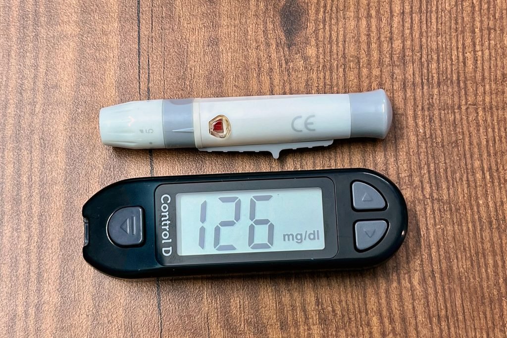
Living successfully with type 2 diabetes requires patients to assume primary responsibility for daily management, supported by their interdisciplinary clinical team.
ROUTINE CLINICAL SURVEILLANCE TIMETABLE
| Frequency | Screening / Diagnostic Action | Clinical Objective |
| Daily | Visual Foot Self-Inspection | Early detection of trauma, blisters, or early pressure ulcers. |
| Every 3 Months (Every 6 Months, if stable) | HbA1c Testing | Tracking glycemic trends; adjusting medication doses as needed |
| Annually | Urine Albumin-to-Creatinine Ratio (uACR) & Serum Creatinine | Early detection of diabetic nephropathy before irreversible eGFR loss occurs |
| Annually | Dilated Eye Examination (Optometrist / Ophthalmologist) | Fundoscopic screening for asymptomatic early microaneurysms and macular edema |
| Annually | Comprehensive Foot Exam (Podiatrist / Clinician) | 10g monofilament, 128Hz tuning fork, and palpation of dorsalis pedis pulses |
| Annually | Complete Fasting Lipid Panel & Blood Pressure Review | Evaluating ASCVD risk; assessing LDL-C and non-HDL-C therapeutic targets |
Continuous Glucose Monitoring (CGM) & The “Time in Range” Paradigm
While quarterly HbA1c measures mean circulating glucose over several months, it cannot capture acute glycemic excursions or dangerous hypoglycemia. Continuous Glucose Monitoring (CGM) systems evaluate dynamic glycemic control using standardized international consensus metrics:
- Time in Range (TIR): The primary goal is to maintain venous or interstitial glucose between 70 and 180 mg/dL (3.9 to 10.0 mmol/L) for >70% of the day. Every 10% increase in TIR correlates with clinically significant reductions in retinopathy and microalbuminuria risk.
- Time Below Range (TBR): Minimize hypoglycemia. Interstitial readings <70 mg/dL (<3.9 mmol/L) must remain under 4% of the day, while severe hypoglycemia <54 mg/dL (<3.0 mmol/L) must remain under 1%.
- Time Above Range (TAR): Glucose readings >180 mg/dL (>10.0 mmol/L) should remain under 25% of the day.
Daily Foot Care Protocol
Peripheral neuropathy combined with microvascular insufficiency means minor skin trauma can rapidly evolve into deep tissue infections or osteomyelitis. Patients must inspect their feet daily using a direct mirror for interdigital erythema, calluses, macerations, blisters, or fissures. Footwear must fit properly without internal seam pressure, and patients should avoid walking barefoot.
Sick-Day Management Rules
Acute illnesses—such as viral gastroenteritis, influenza, or pneumonia—trigger a surge in counter-regulatory stress hormones (cortisol, epinephrine, glucagon), which can precipitate significant hyperglycemia or life-threatening Hyperosmolar Hyperglycemic State (HHS). During illness, patients must:
- Measure blood glucose every 2 to 4 hours.
- Never discontinue basal insulin arbitrarily, even if oral intake is poor, because basal insulin suppression is essential to prevent uncontrolled lipolysis and hepatic gluconeogenesis.
- Maintain oral fluid hydration (at least 200–250 mL of non-caloric fluid per hour while awake).
- Temporarily hold potentially nephrotoxic or risk-elevating medications under clinical guidance using the “SADMAN” protocol: SGLT-2 inhibitors (to prevent euglycemic diabetic ketoacidosis), ACE inhibitors, Diuretics, Metformin (to avoid lactic acidosis during dehydration), ARBs, and NSAIDs.
12. High-Yield Questions about Type 2 Diabetes Mellitus to Ask Your Doctor
Section Summary:
Navigating modern type 2 diabetes requires open, informed communication with healthcare providers. Patients should advocate for comprehensive cardiorenal protection, clarify individualized glycemic and blood pressure targets, assess whether their medication regimen minimizes hypoglycemia risk, understand when to screen for complications, and discuss options for disease remission.
Navigating diabetes care requires patients to play an active, informed role in their management. The following questions are designed to guide meaningful discussions during clinical consultations:
Regarding your Diagnosis & Baseline Risk
- “What is my current cardiovascular and renal risk profile, and what is my formal CKM syndrome stage?”
- “Beyond my HbA1c, what are my urine albumin-to-creatinine ratio (uACR) and my estimated filtration rate (eGFR)?”
- “Am I carrying excess intra-organ or visceral fat, and does my medical history make me an appropriate candidate for structured weight-loss interventions aimed at disease remission?”
Regarding Medication Selection & Organ Protection
- “Do the medications I am currently prescribed merely lower my blood glucose numbers, or do they also provide proven protection against heart failure, stroke, and progressive kidney damage?”
- “Given the latest clinical guidelines, would my health profile benefit from starting an SGLT-2 inhibitor or an incretin-based therapy (GLP-1 RA or dual GIP/GLP-1 RA)?”
- “What is the exact hypoglycemia risk profile of my daily medication regimen, and what signs or symptoms should trigger adjustments?”
Regarding Complication Prevention & Surveillance
- “When was my last comprehensive foot examination using a monofilament and pulse assessment, and am I overdue for a formal evaluation?”
- “Do you have an accredited dilated fundoscopic eye exam report on file for me within the past 12 months?”
- “What specific blood pressure and LDL-cholesterol targets should we aim for to minimize my overall cardiovascular risk?”
Primary Credible Sources
- American Diabetes Association (ADA). (2026). Standards of Care in Diabetes—2026. Diabetes Care, 49(Suppl. 1), S1–S350. https://doi.org/10.2337/dc26-Sint
- Ndumele, C. E., et al. (2023). Cardiovascular-Kidney-Metabolic Health: A Presidential Advisory From the American Heart Association. Circulation, 148(20), 1606–1635. https://doi.org/10.1161/CIR.0000000000001184
- World Health Organization (WHO). (2023). Global Report on Diabetes and Fact Sheet on Diabetes Mellitus. World Health Organization Press, Geneva. https://www.who.int/news-room/fact-sheets/detail/diabetes
- DeFronzo, R. A. (2009). From the Triumvirate to the Ominous Octet: A New Paradigm for the Treatment of Type 2 Diabetes Mellitus. Diabetes, 58(4), 773–795. https://doi.org/10.2337/db09-9028
- Davies, M. J., et al. (2022). Management of Hyperglycemia in Type 2 Diabetes, 2022. A Consensus Report by the American Diabetes Association (ADA) and the European Association for the Study of Diabetes (EASD). Diabetes Care, 45(11), 2753–2786. https://doi.org/10.2337/dci22-0034
- Lean, M. E., et al. (2018). Primary care-led weight management for remission of type 2 diabetes (DiRECT): an open-label, cluster-randomised trial. The Lancet, 391(10120), 541–551. https://doi.org/10.1016/S0140-6736(17)33102-1
- Zinman, B., et al. (EMPA-REG OUTCOME Investigators). (2015). Empagliflozin, Cardiovascular Outcomes, and Mortality in Type 2 Diabetes. New England Journal of Medicine, 373(22), 2117–2128. https://doi.org/10.1056/NEJMoa1504720
- Marso, S. P., et al. (LEADER Steering Committee). (2016). Liraglutide and Cardiovascular Outcomes in Type 2 Diabetes. New England Journal of Medicine, 375(4), 311–322. https://doi.org/10.1056/NEJMoa1603827
- Frias, J. P., et al. (SURPASS-2 Investigators). (2021). Tirzepatide versus Semaglutide Once Weekly in Patients with Type 2 Diabetes. New England Journal of Medicine, 385(6), 503–515. https://doi.org/10.1056/NEJMoa2107519
- Heerspink, H. J. L., et al. (DAPA-CKD Trial Committees). (2020). Dapagliflozin in Patients with Chronic Kidney Disease. New England Journal of Medicine, 383(15), 1436–1446. https://doi.org/10.1056/NEJMoa2024816
- UK Prospective Diabetes Study (UKPDS) Group. (1998). Intensive blood-glucose control with sulphonylureas or insulin compared with conventional treatment and risk of complications in patients with type 2 diabetes (UKPDS 33). The Lancet, 352(9131), 837–853. https://doi.org/10.1016/S0140-6736(98)07019-6
- Taylor, R. (2013). Type 2 Diabetes: Etiology and Reversibility. Diabetes Care, 36(4), 1047–1055. https://doi.org/10.2337/dc12-1805
- Battelino, T., et al. (2019). Clinical Targets for Continuous Glucose Monitoring Data Interpretation: Recommendations From the International Consensus on Time in Range. Diabetes Care, 42(8), 1593–1603. https://doi.org/10.2337/dci19-0028
Featured Image: Markus Winkler / Pexels
© UncutMed — Medical knowledge, precise, truthful, unfiltered. The current article was last modified on September 8, 2026. This article was clinically authored and reviewed under international evidence-based standards. Always consult your personal healthcare physician before modifying any medical therapy.