© UncutMed — Medical knowledge precise, truthful, unfiltered. This article was last reviewed on August 18, 2026. Always consult your healthcare provider for personal medical advice.
⚕️ Medical Disclaimer: This article is for informational and educational purposes only. It does not constitute medical advice, diagnosis, or treatment. Always consult a qualified healthcare professional regarding any medical condition or treatment plan.
Congestive Heart Failure – Clinical Definition

Congestive Heart Failure (CHF) — more accurately termed simply Heart Failure (HF) in modern clinical practice — is a complex clinical syndrome in which the heart is unable to pump sufficient blood to meet the metabolic demands of the body, or can only do so at the cost of abnormally elevated filling pressures. It is not a single disease but rather the final common pathway of numerous cardiac and systemic conditions that progressively impair the heart’s structure or function.
The word “congestive” refers to the accumulation of fluid —congestion— that occurs in the lungs, abdomen, and extremities of the body (such as the ankles) when the failing heart cannot efficiently move blood forward through the circulation. Blood backs up in anatomical structures before reaching the weakened heart, increasing pressure in the veins and capillaries and causing fluid to leak out of blood vessels into surrounding tissues.
It is critically important to understand that heart failure does NOT mean the heart has stopped working. Instead, it means the heart is working inadequately. The heart continues to beat, but its output is insufficient, or its filling pressures are elevated to a degree that causes symptoms and organ dysfunction.
Congestive Heart Failure – Classification
Heart failure is classified into two major subtypes based on the heart’s remaining pumping ability, measured by the ejection fraction (EF) — the percentage of blood ejected from the left ventricle with each heartbeat. Normal ejection fraction is 55–70%:
Heart Failure with Reduced Ejection Fraction (HFrEF)
The Ejection Fraction is below 40%, previously called systolic heart failure. The heart muscle is weakened and contracts poorly, pumping out less blood with each beat. This is the classic form of heart failure most people are familiar with.
Heart Failure with Mildly Reduced Ejection Fraction (HFmrEF)
The Ejection Fraction is 40–49%, a recently recognised intermediate category that shares features of both HFrEF and HFpEF.
Heart Failure with Preserved Ejection Fraction (HFpEF)
The Ejection Fraction is 50% or above, previously called diastolic heart failure. The heart muscle contracts normally and the ejection fraction is preserved, but the muscle has become stiff and cannot relax properly between beats, impairing filling as the heart’s space for blood during relaxation is decreased. Despite normal pumping, the heart cannot accommodate adequate blood volume, leading to elevated pressures and the same congestion symptoms as HFrEF.
Symptoms of Congestive Heart Failure
Symptoms in heart failure arise from two fundamental physiological derangements: reduced forward flow of blood to the body’s organs, and backward congestion of blood behind (meaning that the blood stays in tissues longer before returning to the heart through circulation) the failing heart. Understanding which mechanism drives each symptom makes them far easier to comprehend and recognise.
- Dyspnoea — Breathlessness/Difficulty breathing. Breathlessness is the cardinal symptom of heart failure and the one that most commonly brings patients to medical attention. It occurs because the failing left ventricle cannot pump blood forward efficiently, causing it to back up into the pulmonary veins and capillaries of the lungs. As pulmonary capillary pressure rises, fluid leaks from the capillaries into the air spaces of the lungs — a condition called pulmonary oedema. The presence of fluid in the lungs reduces their compliance, increases the work of breathing, and impairs gas exchange, producing the sensation of breathlessness. Breathlessness in heart failure presents in several characteristic patterns: 1. Exertional dyspnoea, breathlessness on physical activity — is typically the earliest symptom, reflecting the heart’s inability to increase its output sufficiently to meet the demands of exercise. 2. Orthopnoea — breathlessness when lying flat — occurs because the supine (lying flat on your back) position redistributes fluid from the legs and abdomen into the pulmonary circulation, further increasing pulmonary venous pressure. Patients characteristically sleep with multiple pillows to keep themselves upright and relieve this symptom. 3. Paroxysmal Nocturnal Dyspnoea (PND) — episodes of sudden severe breathlessness that wake the patient from sleep, typically 1 to 2 hours after lying down. The patient wakes gasping for air, sits upright, and gradually improves over 15 to 30 minutes. PND reflects the same mechanism as orthopnoea but in a more dramatic acute form, as fluid redistribution overwhelms the pulmonary circulation during sleep.
- Fatigue and Exercise Intolerance. Profound fatigue is one of the most debilitating symptoms of heart failure and is driven by reduced cardiac output — the failing heart simply cannot pump enough blood, so it cannot deliver adequate oxygen and nutrients to skeletal muscles during activity. Additionally, chronic neurohormonal activation in heart failure — particularly elevated catecholamines — causes skeletal muscle wasting and metabolic dysfunction, further impairing exercise capacity even when cardiac output is only mildly reduced.
- Peripheral Oedema — Fluid Retention. Swelling of the ankles, legs, and, in severe cases, the abdomen and genitals is a hallmark of right-sided heart failure or biventricular failure. When the right ventricle fails, blood backs up into the systemic venous circulation, raising venous pressure in the peripheral capillaries. This elevated hydrostatic pressure drives fluid out of capillaries into the interstitial tissues, producing the characteristic pitting oedema of heart failure — so named because pressing a finger into the swollen tissue leaves a temporary indentation (pit). Additionally, the kidneys respond to reduced cardiac output by activating the renin-angiotensin-aldosterone system (RAAS), causing sodium and water retention that further exacerbates fluid accumulation.
- Ascites. In severe right-sided heart failure, elevated venous pressure in the portal circulation (the blood supply to the liver and intestines) causes fluid to accumulate within the abdominal cavity — a condition known as ascites. This produces abdominal distension, discomfort, and a sensation of early satiety as the enlarged abdomen compresses the stomach.
- Nocturia — Frequent urination during the night. During the day, reduced cardiac output causes the kidneys to retain sodium and water. However, when the patient lies down at night, venous return to the heart increases, temporarily improving cardiac output and renal perfusion, prompting the kidneys to excrete the excess fluid accumulated during the day as urine. This explains the characteristic pattern of heart failure patients needing to urinate frequently during the night despite reduced urine output during the day.
- Cardiac Cachexia. In advanced heart failure, severe unintentional weight loss and muscle wasting — termed cardiac cachexia — can develop. This results from chronically reduced intestinal perfusion impairing nutrient absorption, elevated inflammatory cytokines promoting catabolism, increased metabolic demand from the work of laboured breathing, and reduced appetite from abdominal congestion and medication side effects. Cardiac cachexia is a marker of advanced disease and carries a poor prognosis.
- Cognitive Impairment. Reduced cerebral perfusion- meaning that the heart doesn’t supply the brain with adequate blood– in heart failure can cause difficulty concentrating, memory impairment, confusion, and, in severe cases, delirium, particularly in elderly patients. Cerebral hypoperfusion activates compensatory mechanisms that may temporarily maintain brain blood flow at the expense of other organs but eventually proves insufficient in advanced disease.
- Reduced Urine Output (Oliguria). As cardiac output falls, renal perfusion decreases, activating neurohormonal compensatory mechanisms that cause the kidneys to retain sodium and water and reduce urine production. Severely reduced urine output (oliguria) in heart failure indicates significant renal hypoperfusion and is a marker of haemodynamic compromise.
Signs of Congestive Heart Failure
Signs are objective findings detected by a clinician during physical examination. In heart failure, they reflect the same pathophysiological mechanisms as symptoms, backward congestion and reduced forward flow, but are identified through clinical assessment rather than patient report.
- Elevated Jugular Venous Pressure (JVP). One of the most important and informative signs in cardiovascular medicine. The jugular veins in the neck are continuous with the right atrium and reflect right-sided filling pressures. In right heart failure, elevated right atrial pressure causes the jugular veins to become visibly distended — a raised JVP. Clinicians estimate right atrial pressure by measuring the height of the venous pulsation above the sternal angle with the patient reclined at 45 degrees. A Jugular Venous Pressure raised more than 3–4 cm above the sternal angle is abnormal and indicates elevated right-sided filling pressures.
- Third Heart Sound (S3 Gallop). Normally, two heart sounds are heard — S1 (closure of the mitral and tricuspid valves at the start of systole) and S2 (closure of the aortic and pulmonary valves at the end of systole). In heart failure, a third heart sound — S3 — can be heard in early diastole, immediately after S2. It is produced by the sudden deceleration of blood flowing rapidly into a dilated, poorly compliant ventricle. The presence of S3 in an adult is a highly specific sign of elevated ventricular filling pressures and significant cardiac dysfunction.
- Displaced Apex Beat. The apex beat is the point of maximum cardiac impulse felt on the chest wall. Normally located in the fifth intercostal space in the midclavicular line, in heart failure, the dilated left ventricle pushes the apex beat downward and outward — toward the axilla. A displaced apex beat indicates cardiomegaly (enlarged heart) and is an important clinical sign of structural cardiac remodelling.
- Bibasal Crackles (Pulmonary Crepitations). Auscultation of the lungs in heart failure reveals fine crackling sounds — crepitations — heard at the lung bases bilaterally. These are produced by the opening of small airways and alveoli that have been collapsed or filled with fluid during expiration. Bibasal crackles in the appropriate clinical context are a reliable sign of pulmonary congestion and indicate elevated left-sided filling pressures.
- Hepatomegaly and Hepatojugular Reflux. Congestion of the hepatic veins in right heart failure causes the liver to become enlarged and tender — hepatomegaly. Pressing firmly on the congested liver increases venous return to the right heart, which — in the presence of elevated right-sided pressures — produces a visible rise in the jugular venous pressure. This is called the hepatojugular reflux and is a useful confirmatory sign of right heart failure and elevated central venous pressure.
- Peripheral Oedema. Clinically detected as pitting oedema of the ankles and legs — bilateral, symmetrical, and gravitationally dependent. In bed-bound patients, fluid accumulates in the sacral region rather than the ankles, making sacral oedema an important sign to check in hospitalised patients. Severe oedema can extend up the legs to the thighs and abdomen.
- Tachycardia. The failing heart compensates for reduced stroke volume by increasing heart rate — a response mediated by activation of the sympathetic nervous system and elevated catecholamines. Persistent resting tachycardia in heart failure reflects ongoing sympathetic activation and haemodynamic compromise. Paradoxically, this compensatory tachycardia worsens heart failure over time by increasing myocardial oxygen demand and impairing diastolic filling time.
- Hypotension and Narrow Pulse Pressure. In advanced heart failure, reduced cardiac output causes a fall in systolic blood pressure. The pulse pressure — the difference between systolic and diastolic blood pressure — narrows as cardiac output falls, reflecting reduced stroke volume. A systolic blood pressure below 90 mmHg in a heart failure patient indicates cardiogenic shock — a life-threatening emergency.
- Cardiac Murmurs. Functional mitral or tricuspid regurgitation murmurs are commonly heard in heart failure. As the ventricles dilate, the valve annuli stretch, preventing complete leaflet coaptation and causing regurgitation — blood leaking backward through the valve during systole. These functional murmurs worsen heart failure by reducing forward cardiac output and further elevating filling pressures.
Congestive Heart Failure – Causes

Heart failure is the end result of any condition that damages, weakens, or chronically overloads the heart muscle. The most common causes globally are:
- Coronary Artery Disease and Myocardial Infarction. The most common cause of heart failure in the developed world. Atherosclerotic narrowing of the coronary arteries chronically deprives the heart muscle of oxygen, causing ischaemic cardiomyopathy — progressive weakening and scarring of the myocardium. A myocardial infarction causes acute death of a region of heart muscle, permanently replacing contractile tissue with non-functional scar tissue. Depending on the size and location of the infarction, the resulting loss of functional myocardium can dramatically reduce ejection fraction and precipitate heart failure. For a complete guide on coronary artery disease, read the article on UncutMed.
- Hypertension. Chronically elevated blood pressure forces the left ventricle to work against increased resistance to pump blood forward with every heartbeat — a condition called pressure overload. Initially, the ventricle compensates by thickening its walls (concentric hypertrophy), maintaining normal cardiac output at the cost of increased wall stress and oxygen demand. Over years, this compensatory hypertrophy becomes maladaptive — the thickened muscle becomes stiff, impairing diastolic filling (HFpEF), and eventually the overstressed myocardium dilates and weakens, leading to HFrEF. Hypertension is the leading cause of HFpEF globally.
- Cardiomyopathies. Primary diseases of the heart muscle that alter its structure and function. There are various types of cardiomyopathies. Dilated Cardiomyopathy — the ventricle dilates and weakens, reducing ejection fraction. Causes include viral myocarditis, alcohol toxicity, certain medications (e.g., chemotherapy agents such as anthracyclines), genetic mutations, and idiopathic (unknown) causes. Hypertrophic Cardiomyopathy — a genetic mutation that causes abnormal thickening of the ventricular walls, impairing diastolic filling and causing outflow obstruction. Restrictive Cardiomyopathy — infiltration of the myocardium with abnormal material (e.g., amyloid protein in cardiac amyloidosis, iron in haemochromatosis) makes the ventricle rigid and non-compliant, severely impairing filling.
- Valvular Heart Disease. Diseased heart valves impose abnormal pressure or volume loads on the cardiac chambers, leading to various valve disorders. Aortic Stenosis: narrowing of the aortic valve creates severe pressure overload on the left ventricle, driving compensatory hypertrophy that eventually leads to heart failure. Mitral Regurgitation — a leaking mitral valve allows blood to flow backward into the left atrium during systole, creating volume overload on the left ventricle that over time causes dilatation and systolic dysfunction. Aortic Regurgitation — a leaking aortic valve causes massive volume overload on the left ventricle as blood regurgitates back from the aorta during diastole, eventually leading to ventricular dilatation and failure.
- Arrhythmias. Atrial Fibrillation (AF) is both a cause and consequence of heart failure. Sustained rapid heart rates in uncontrolled AF reduce ventricular filling time, decrease cardiac output, and over months cause tachycardia-mediated cardiomyopathy — a reversible form of heart failure that can recover if the arrhythmia is controlled.
- Congenital heart disease — structural abnormalities present from birth that impose chronic haemodynamic burdens on the heart.
- Myocarditis — acute inflammation of the myocardium, most commonly viral, that can cause acute or chronic heart failure.
- Pericardial disease — constrictive pericarditis prevents normal cardiac filling, mimicking and eventually causing heart failure physiology.
- High output states — conditions such as severe anaemia, thyrotoxicosis, and arteriovenous fistulae demand chronically elevated cardiac output that eventually exhausts even a structurally normal heart.
- Cardiotoxic medications — certain chemotherapy agents, alcohol, cocaine, and methamphetamine directly damage the myocardium.
Risk Factors for Congestive Heart Failure
Risk factors for heart failure largely mirror those for coronary artery disease, reflecting the dominant role of CAD in its aetiology, but include several additional important contributors:
- Coronary Artery Disease — the single most important risk factor for heart failure.
- Hypertension — particularly long-standing, poorly controlled hypertension.
- Diabetes Mellitus — independently increases heart failure risk through diabetic cardiomyopathy — direct glucose-mediated myocardial damage — in addition to its role in accelerating CAD.
- Age — the prevalence of heart failure doubles with each decade after age 45, reflecting the cumulative burden of risk factors and age-related changes in cardiac structure and function.
- Male Sex — men develop HFrEF more commonly; women are more prone to HFpEF.
- Family History — genetic predisposition plays a role, particularly in cardiomyopathies.
- Previous Myocardial Infarction — the most powerful single predictor of subsequent heart failure.
- Valvular Heart Disease — particularly aortic stenosis and mitral regurgitation.
- Atrial Fibrillation — bidirectional relationship with heart failure.
- Chronic Kidney Disease — cardiorenal syndrome creates a vicious cycle of worsening cardiac and renal function.
- Sleep Apnoea — causes nocturnal hypoxia, pulmonary hypertension, and right heart strain.
- Alcohol and Substance Use — dose-dependent cardiotoxicity.
- Chemotherapy Exposure — particularly anthracyclines (doxorubicin) and trastuzumab.
- Sedentary Lifestyle — independently associated with increased heart failure risk.
- Smoking — accelerates atherosclerosis and directly damages the myocardium.
Stages of Heart Failure — The ACC/AHA Classification
The American College of Cardiology and American Heart Association classify heart failure into four stages — A through D — that describe the progressive evolution of the disease from risk to end-stage. This staging system is distinct from the NYHA functional classification (which describes symptom severity) and is important because it emphasises that heart failure prevention and intervention should begin before symptoms develop.
Stage A — At Risk for Heart Failure
Definition: The patient has risk factors for heart failure but no structural heart disease and no symptoms.
Who this includes: Patients with hypertension, diabetes, obesity, coronary artery disease, metabolic syndrome, exposure to cardiotoxic medications, family history of cardiomyopathy, or a history of rheumatic fever.
Physiological rationale: At this stage, the myocardium has not yet sustained significant damage. The heart’s structure and function remain normal. However, the presence of risk factors means that pathological processes — endothelial injury, neurohormonal activation, myocardial stress — are already operating at a subclinical level that will progressively damage cardiac structure if left unaddressed.
Management focus: Aggressive risk factor modification — blood pressure control, diabetes management, smoking cessation, weight loss, and treatment of dyslipidaemia — to prevent progression to structural heart disease.
Stage B — Pre-Heart Failure (Structural Heart Disease Without Symptoms)
Definition: The patient has developed structural heart disease — such as reduced ejection fraction, left ventricular hypertrophy, or valvular abnormalities — but has never experienced symptoms of heart failure.
Who this includes: Patients with a previous myocardial infarction, reduced ejection fraction on echocardiogram, left ventricular hypertrophy on ECG or echo, or significant valvular disease — all without symptoms.
Physiological rationale: At this stage, the myocardium has sustained structural changes — loss of contractile tissue, pathological hypertrophy, or remodelling — but compensatory mechanisms (neurohormonal activation, ventricular dilatation, tachycardia) are sufficient to maintain cardiac output at rest. Symptoms do not yet occur because the heart can still meet baseline metabolic demands. However, these compensatory mechanisms are themselves progressively harmful — ventricular remodelling and neurohormonal activation accelerate further structural deterioration.
Management focus: Medications proven to prevent progression to symptomatic heart failure — particularly ACE inhibitors or ARBs and beta-blockers in patients with reduced EF — alongside continued risk factor management. Valvular disease may require surgical correction at this stage.
Stage C — Symptomatic Heart Failure
Definition: The patient has structural heart disease and current or prior symptoms of heart failure — breathlessness, fatigue, reduced exercise tolerance, fluid retention.
Who this includes: The majority of patients diagnosed with clinical heart failure. This stage encompasses patients ranging from those with mild exertional symptoms to those with severe functional limitation.
Physiological rationale: Compensatory mechanisms are no longer sufficient to maintain cardiac output without the cost of elevated filling pressures and fluid congestion. The neurohormonal activation that initially compensated for reduced cardiac output — sympathetic activation, RAAS activation, ADH release — now becomes chronically maladaptive, causing progressive ventricular remodelling, sodium and water retention, vasoconstriction, and myocardial toxicity from elevated catecholamines.
Management focus: Comprehensive medical therapy targeting the maladaptive neurohormonal pathways — ACE inhibitors/ARBs/ARNI, beta-blockers, mineralocorticoid receptor antagonists, SGLT-2 inhibitors — alongside diuretics for symptom relief, device therapy (ICD, CRT) where indicated, and lifestyle modification. The goal is symptom control, prevention of hospitalisation, and reduction of mortality.
Stage D — Advanced Heart Failure
Definition: The patient has severe, refractory heart failure symptoms at rest or with minimal exertion despite optimal medical therapy. This represents end-stage heart failure.
Who this includes: Patients with frequent hospitalisations for heart failure decompensation, those who cannot be weaned from intravenous inotropic support, and those with severely reduced cardiac output causing end-organ damage — cardiorenal syndrome, cardiac cachexia, or cardiogenic shock.
Physiological rationale: At this stage, the myocardium is so extensively damaged that no degree of neurohormonal compensation or medical therapy can maintain adequate cardiac output. Organ perfusion is chronically insufficient, driving a cycle of progressive multi-organ dysfunction. The compensatory mechanisms that were initially protective — RAAS activation, sympathetic stimulation — are now so chronically activated that they are themselves causing organ damage.
Management focus: Advanced therapies — continuous intravenous inotropic support, mechanical circulatory support devices (left ventricular assist device — LVAD), cardiac transplantation, or palliative care with a focus on symptom management and quality of life for patients who are not candidates for advanced interventions.
Diagnostic Tests for Congestive Heart Failure
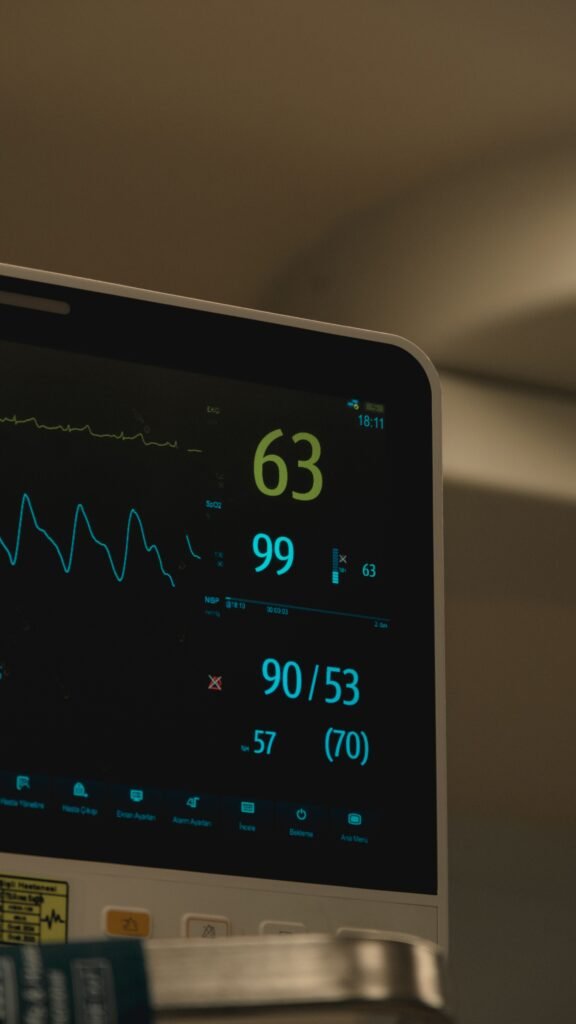
The diagnosis of heart failure is clinical — based on symptoms and signs — but is confirmed and characterised by a combination of investigations:
Blood Tests
BNP and NT-proBNP (Natriuretic Peptides): The most important blood tests for diagnosing heart failure. BNP (B-type natriuretic peptide) and its inactive fragment NT-proBNP are hormones secreted by the ventricular myocardium in response to elevated wall stress and volume overload. Elevated levels strongly support a diagnosis of heart failure and correlate with disease severity. They are also used to monitor treatment response and guide therapy — falling natriuretic peptide levels indicate improving haemodynamics.
Full Blood Count: Anaemia — a common finding in heart failure — worsens symptoms by reducing oxygen delivery to tissues and increasing cardiac workload. Polycythaemia may indicate chronic hypoxia.
Renal Function and Electrolytes: Chronic renal impairment is both a cause and consequence of heart failure. Electrolyte disturbances — particularly hypokalaemia and hyponatraemia — are common in heart failure and are worsened by diuretic therapy. Hyponatraemia in heart failure is a marker of severe neurohormonal activation and carries a poor prognosis.
Liver Function Tests: Hepatic congestion from right heart failure causes elevated bilirubin and liver enzymes — a pattern sometimes called congestive hepatopathy or cardiac cirrhosis in severe chronic cases.
Thyroid Function Tests: Both hypothyroidism and hyperthyroidism can cause or exacerbate heart failure and must be excluded in all new presentations.
Fasting Glucose and HbA1c: To identify diabetes as a contributing cause and guide management.
Iron Studies: Iron deficiency is extremely common in heart failure — present in up to 50% of patients — and is independently associated with worse symptoms and outcomes regardless of whether anaemia is present. Intravenous iron supplementation has been shown to improve symptoms and reduce hospitalisations.
Electrocardiogram (ECG)
A 12-lead ECG is performed in all patients with suspected heart failure. While not diagnostic of heart failure itself, it provides critical information about:
- Evidence of previous myocardial infarction (pathological Q waves)
- Left ventricular hypertrophy (voltage criteria)
- Atrial fibrillation or other arrhythmias
- Left bundle branch block — important for identifying patients who may benefit from cardiac resynchronisation therapy (CRT)
- QT interval prolongation — relevant for medication safety
A completely normal ECG makes significant cardiac dysfunction unlikely and should prompt reconsideration of the diagnosis.
Chest X-Ray
The chest X-ray in heart failure characteristically shows:
- Upper lobe venous diversion — pulmonary veins in the upper lobes become more prominent than lower lobe veins as elevated pulmonary venous pressure diverts flow upward.
- Cardiomegaly — cardiac silhouette occupying more than 50% of the thoracic diameter on a PA (posteroanterior) film.
- Kerley B lines — horizontal lines at the lung bases representing fluid-filled interlobular septa.
- Perihilar haziness — a bat-wing pattern of pulmonary oedema in severe cases.
- Pleural effusions — fluid collections in the pleural space, typically bilateral and right-sided predominant.
Echocardiogram
The cornerstone investigation for heart failure provides a comprehensive assessment of cardiac structure and function. The echocardiogram measures:
- Ejection fraction — the most important single parameter, determining HFrEF vs HFpEF.
- Ventricular size and wall thickness — identifying dilatation or hypertrophy.
- Wall motion abnormalities — regions of the ventricle contracting poorly due to ischaemia or infarction.
- Diastolic function — tissue Doppler and other parameters assess ventricular relaxation and filling pressures.
- Right ventricular function and pulmonary artery pressure — assessing for right heart failure and pulmonary hypertension.
- Pericardial effusion — fluid around the heart.
Cardiac MRI
The gold standard for myocardial tissue characterisation — providing the most accurate assessment of ventricular volumes, ejection fraction, and myocardial mass. Crucially, cardiac MRI with gadolinium contrast can identify:
- Late gadolinium enhancement — areas of myocardial fibrosis and scar from previous infarction or cardiomyopathy.
- Myocardial inflammation — in myocarditis.
- Infiltrative disease — cardiac amyloidosis, sarcoidosis, haemochromatosis.
- Myocardial viability — distinguishing hibernating viable myocardium from irreversible scar — critical for deciding whether revascularisation will improve cardiac function.
Coronary Angiography
Performed to identify or exclude Coronary Artery Disease as the underlying cause of heart failure — particularly important in patients presenting with new heart failure and risk factors for CAD. Identifying significant coronary disease is crucial because revascularisation (PCI or CABG) may restore blood flow to hibernating myocardium and improve ejection fraction.
Cardiopulmonary Exercise Testing (CPET)
A specialised test measuring oxygen consumption and carbon dioxide production during maximal exercise on a treadmill or bicycle. The peak VO2 — maximum oxygen uptake — is the gold standard measure of functional capacity in heart failure and is used to objectively quantify exercise limitation, guide prognosis, and, in advanced heart failure, determine candidacy for cardiac transplantation.
Right Heart Catheterisation
Invasive measurement of intracardiac pressures and cardiac output using a catheter advanced through a vein into the right heart and pulmonary artery. It provides the most accurate haemodynamic assessment of heart failure severity — measuring right atrial pressure, pulmonary artery pressure, pulmonary capillary wedge pressure (reflecting left atrial pressure), and cardiac output directly. Essential in the evaluation of patients being considered for advanced therapies such as LVAD or cardiac transplantation.
Treatment of Congestive Heart Failure

Photo: Tima Miroshnichenko / Pexels
Treatment of heart failure aims to relieve symptoms, prevent hospitalisation, slow down disease progression, reverse pathological cardiac remodelling, and reduce mortality. It is divided into pharmacological and non-pharmacological approaches, with device and surgical therapy for selected patients.
Pharmacological Treatment
Modern heart failure pharmacotherapy has been revolutionised over the past decade by the emergence of four drug classes that together form the cornerstone of HFrEF treatment — termed the “four pillars” of heart failure therapy by current ESC guidelines.
Pillar 1 — Angiotensin-converting enzyme (ACE) Inhibitors / Angiotensin II receptor blockers (ARBs) / Angiotensin Receptor-Neprilysin Inhibitors (ARNI)
ACE inhibitors (e.g., ramipril, enalapril) and ARBs (e.g., candesartan, valsartan) block the renin-angiotensin-aldosterone system — the primary neurohormonal pathway driving fluid retention, vasoconstriction, and maladaptive cardiac remodelling in heart failure. By blocking angiotensin II production or action, these drugs reduce preload and afterload, promote natriuresis, and critically reduce ventricular remodelling — slowing or reversing the progressive dilatation and dysfunction of the failing heart. They reduce mortality in HFrEF by approximately 20–25%.
The ARNI sacubitril/valsartan (Entresto) combines an ARB with a neprilysin inhibitor that amplifies the beneficial effects of natriuretic peptides. It has been shown to reduce cardiovascular mortality and heart failure hospitalisation by 20% compared to ACE inhibitor therapy alone and is now the preferred first-line agent in HFrEF patients who can tolerate it.
Pillar 2 — Beta-Blockers
Beta-blockers (bisoprolol, carvedilol, metoprolol succinate) counteract the chronic sympathetic nervous system activation that characterises heart failure. While for a short period of time the sympathetic activation compensates for reduced cardiac output by increasing heart rate and contractility, chronically elevated catecholamines are cardiotoxic — causing myocyte apoptosis, arrhythmias, and progressive ventricular remodelling. Beta-blockers reduce heart rate, decrease myocardial oxygen demand, protect against arrhythmic death, and over months to years allow the failing ventricle to recover function — a phenomenon called reverse remodelling. They reduce all-cause mortality in HFrEF by approximately 34%
Pillar 3 — Mineralocorticoid Receptor Antagonists (MRAs)
Spironolactone and eplerenone block aldosterone receptors in the kidney and myocardium. Aldosterone — chronically elevated in heart failure through RAAS activation — causes renal sodium retention, potassium wasting, and directly promotes myocardial and vascular fibrosis. MRAs reduce fluid retention, prevent hypokalaemia from other diuretics, and crucially reduce myocardial fibrosis and pathological remodelling. They reduce mortality in HFrEF by approximately 30% and are recommended for all patients with HFrEF and an eGFR above 30 mL/min who can maintain safe potassium levels.
Pillar 4 — SGLT-2 Inhibitors (sodium-glucose co-transporter 2)
The most exciting recent advance in heart failure pharmacotherapy. Originally developed as diabetes medications, SGLT-2 inhibitors (dapagliflozin, empagliflozin) were discovered to have profound and independent cardiovascular benefits. They reduce heart failure hospitalisations and cardiovascular mortality in both HFrEF and HFpEF — the only drug class proven to reduce mortality in HFpEF. Their mechanisms in heart failure are multiple and not fully elucidated, but include osmotic diuresis reducing preload, natriuresis, reduced inflammation and oxidative stress, improved myocardial energetics, and direct anti-fibrotic effects on the myocardium. They are now recommended for all patients with HFrEF regardless of diabetic status.
Diuretics
Loop diuretics (furosemide, bumetanide, torasemide) are the most effective agents for rapidly relieving congestion — the breathlessness, oedema, and fluid overload that cause the most distressing symptoms of heart failure. They act on the loop of Henle, an anatomical structure in the kidneys, blocking sodium and water reabsorption and promoting rapid diuresis. While diuretics provide dramatic symptomatic relief, they do not reduce mortality and must be used at the lowest effective dose to avoid volume depletion, electrolyte disturbances, and neurohormonal activation.
Ivabradine
Ivabradine selectively reduces heart rate by blocking the funny current (they are named “funny” because they do the opposite of most heart channels: they open up and let positive sodium and potassium ions in when the cell becomes more negative, a state called hyperpolarization) in the sinoatrial node — without affecting blood pressure or myocardial contractility. It is indicated in HFrEF patients who remain symptomatic with a resting heart rate above 70 beats per minute despite optimal beta-blocker therapy. Elevated resting heart rate in heart failure indicates ongoing sympathetic activation and is independently associated with worse outcomes. Reducing heart rate with ivabradine prolongs diastolic filling time, reduces myocardial oxygen demand, and improves cardiac output.
Digoxin
One of the oldest cardiac medications, digoxin inhibits the sodium-potassium ATPase pump in myocardial cells, increasing intracellular calcium and enhancing contractility. In heart failure, it also has vagotonic effects that slow the ventricular rate in atrial fibrillation. While digoxin reduces heart failure hospitalisations and improves symptoms, it has not been shown to reduce mortality and has a narrow therapeutic window (meaning that the dose gap between therapeutic and toxic levels is small), with toxic levels causing dangerous arrhythmias. Its use is now largely limited to patients with HFrEF and concomitant atrial fibrillation not controlled by other agents.
Anticoagulation
Patients with heart failure and concurrent atrial fibrillation require anticoagulation to prevent cardioembolic stroke — preferably with direct oral anticoagulants (DOACs) such as apixaban or rivaroxaban over warfarin due to superior safety profiles. Anticoagulation is not routinely recommended in heart failure patients in sinus rhythm unless other indications exist, as the benefit does not outweigh the bleeding risk in this population.
Device Therapy
Implantable Cardioverter Defibrillator (ICD)
Sudden cardiac death from ventricular arrhythmias is the leading cause of mortality in patients with HFrEF. An ICD continuously monitors cardiac rhythm and delivers a life-saving electrical shock to terminate ventricular fibrillation or ventricular tachycardia. It is recommended for patients with HFrEF and an ejection fraction below 35% who are on optimal medical therapy and have a life expectancy of more than one year — reducing sudden cardiac death risk by approximately 50%.
Cardiac Resynchronisation Therapy (CRT)
In many heart failure patients, abnormal electrical conduction — particularly left bundle branch block — causes the two ventricles to contract in a dyssynchronous (uncoordinated) manner, further reducing cardiac efficiency. CRT uses a specialised pacemaker with leads placed in both ventricles to restore synchronous contraction, improving mechanical efficiency and cardiac output. In appropriately selected patients, CRT improves symptoms, increases ejection fraction, reverses ventricular remodelling, reduces hospitalisations, and reduces mortality. Many patients receive a combined CRT-D device providing both resynchronisation and defibrillation.
Surgical and Advanced Therapies
Coronary Revascularisation
In patients with ischaemic cardiomyopathy — heart failure caused by coronary artery disease — regions of the myocardium may be chronically ischaemic but still viable (hibernating myocardium). These regions contract poorly due to chronic ischaemia but retain the potential to recover function if blood flow is restored through PCI (Percutaneous Coronary Intervention, commonly known as coronary angioplasty with stenting) or CABG (Coronary Artery Bypass Grafting, commonly known as bypass surgery). Identifying and revascularising viable hibernating myocardium can significantly improve ejection fraction and functional status of the heart.
Valvular Intervention
Correction of the underlying valvular lesion causing or exacerbating heart failure — whether through surgical repair/replacement or transcatheter techniques — can dramatically improve cardiac function. Transcatheter aortic valve implantation (TAVI) has revolutionised the treatment of aortic stenosis in high-surgical-risk patients. The MitraClip procedure allows percutaneous reduction of mitral regurgitation in selected patients with functional mitral regurgitation complicating heart failure.
Left Ventricular Assist Device (LVAD)
A mechanical pump surgically implanted to assist the failing left ventricle in pumping blood to the aorta. LVADs are used as a bridge to cardiac transplantation in patients awaiting a donor heart, or as destination therapy in patients who are not transplant candidates. Modern LVADs significantly improve survival and quality of life in Stage D heart failure — one-year survival with LVAD approaches 80% in carefully selected patients.
Cardiac Transplantation
The gold standard treatment for end-stage heart failure refractory to all other therapies. Cardiac transplantation offers five-year survival of approximately 70–75% in carefully selected recipients. It is limited by the severe shortage of donor organs and requires lifelong immunosuppression. Candidates are carefully selected through comprehensive evaluation, including cardiopulmonary exercise testing, right heart catheterisation, and multidisciplinary review.
Prevention of Congestive Heart Failure

The most effective strategy against heart failure is preventing it from developing in the first place. Since the majority of heart failure cases result from conditions that are themselves preventable or modifiable, a significant proportion of the global heart failure burden is avoidable.
As with coronary artery disease, lifestyle modifications are not merely preventive measures — they are clinically proven therapeutic interventions that reduce disease progression at every stage. For patients already diagnosed with heart failure, the following measures form an essential and active component of management, not optional additions to medication.
Control Blood Pressure Rigorously
Hypertension is the most important modifiable risk factor for heart failure — particularly HFpEF. Maintaining blood pressure below 130/80 mmHg through lifestyle modification and medication dramatically reduces the risk of developing heart failure and slows progression in those already affected. Every 10 mmHg reduction in systolic blood pressure reduces heart failure risk by approximately 28%.
Prevent and Manage Coronary Artery Disease through lifestyle modifications
Since Coronary Artery Disease (CAD) is the leading cause of heart failure, all measures that prevent or treat CAD — lowering the lipid levels in the bloodstream, antiplatelet therapy, smoking cessation, diabetes management — directly reduce heart failure incidence. Prompt and effective treatment of myocardial infarction — particularly rapid primary PCI — minimises the extent of myocardial damage and reduces the risk of post-infarction heart failure.
Achieve and Maintain a Healthy Weight
Obesity independently doubles the risk of heart failure through multiple mechanisms — haemodynamic overload, metabolic dysfunction, sleep apnoea, and direct lipotoxic effects on the myocardium. Weight loss in obese patients reduces left ventricular mass, improves diastolic function, and can reverse early structural cardiac changes.
Exercise Regularly
Regular aerobic exercise reduces blood pressure, improves insulin sensitivity, aids weight management, and has direct beneficial effects on cardiac structure and function — reducing left ventricular mass and improving diastolic function. In patients with established heart failure, supervised exercise training through cardiac rehabilitation reduces hospitalisations and improves quality of life and functional capacity.
Eliminate Alcohol and Avoid Cardiotoxins
Alcohol is directly cardiotoxic in a dose-dependent manner — chronic heavy alcohol consumption causes alcoholic cardiomyopathy, a potentially reversible form of dilated cardiomyopathy if alcohol is eliminated early. Complete abstinence in alcohol-related cardiomyopathy can lead to significant recovery of ejection fraction. Cocaine and methamphetamine cause acute coronary spasm, myocarditis, and cardiomyopathy and must be avoided entirely.
Optimise Diabetes Management
Precise and regular glycaemic control reduces microvascular complications and the direct cardiotoxic effects of hyperglycaemia on the myocardium. SGLT-2 (sodium-glucose co-transporter 2) inhibitors and GLP-1 (glucagon-like peptide-1) receptor agonists — beyond their glucose-lowering effects — have proven cardiovascular protective properties and should be prioritised in diabetic patients with or at risk of heart failure.
Treat Sleep Apnoea
Continuous Positive Airway Pressure (CPAP) therapy for obstructive sleep apnoea reduces nocturnal hypoxia, lowers blood pressure, reduces pulmonary hypertension, and improves right ventricular function — all of which reduce heart failure risk and improve outcomes in those with established disease.
Monitor and Treat Atrial Fibrillation
Prompt rate and rhythm control of atrial fibrillation prevents tachycardia-mediated cardiomyopathy — a reversible form of heart failure that can recover completely with successful AF treatment.
Regular Cardiovascular Screening
Routine medical check-ups allow early identification of hypertension, diabetes, obesity, dyslipidaemia, and early structural cardiac changes — enabling intervention before heart failure develops. Patients with Stage A or B heart failure benefit from echocardiographic surveillance and preventive medical therapy.
Living With Congestive Heart Failure — Life and Prognosis

A diagnosis of heart failure is a significant life event — but with modern therapy, the outlook has improved dramatically over the past two decades. Understanding what to expect allows patients to make informed decisions and engage actively in their own care.
Prognosis
Heart failure prognosis varies enormously depending on the underlying cause, ejection fraction, stage, comorbidities, and response to therapy. General prognostic benchmarks:
- Five-year mortality across all heart failure stages is approximately 50% — comparable to many common cancers — though this figure encompasses a vast spectrum from mild Stage C to end-stage Stage D disease, so it may be misleading.
- HFrEF treated with optimal modern therapy (all four pillars) has a significantly improved prognosis compared to historical data — some studies show mortality reductions of over 60% with quadruple therapy.
- HFpEF historically had fewer proven therapies, but SGLT-2 inhibitors have now demonstrated mortality benefit in this group of patients.
- Reversible causes — tachycardia-mediated cardiomyopathy, alcohol cardiomyopathy, peripartum cardiomyopathy — can see substantial or complete recovery of ejection fraction with appropriate treatment.
- Adverse prognostic markers include severe reduction in ejection fraction, elevated natriuretic peptides, hyponatraemia, renal impairment, cardiac cachexia, recurrent hospitalisations, and low peak VO2 on exercise testing.
Daily Life Management
Activity and Exercise
Contrary to historical advice to rest, current guidelines strongly encourage regular physical activity in stable heart failure. Supervised cardiac rehabilitation programmes incorporating structured aerobic exercise improve functional capacity, quality of life, and reduce hospitalisation rates. Patients should pace themselves, avoid exertion in extreme levels of temperature, and rest when symptomatic.
Fluid and Sodium Restriction
Sodium restriction to below 2 grams per day reduces fluid retention and the diuretic dose required to maintain euvolaemia. In advanced heart failure, fluid restriction to 1.5–2 litres per day may be recommended — excessive fluid intake overwhelms the diuretic regimen and precipitates decompensation.
Daily Weight Monitoring
Patients with heart failure should weigh themselves every morning after urinating and before eating. A weight gain of more than 2 kg in 2 days or 3 kg in 1 week indicates fluid accumulation and warrants urgent contact with their medical team for diuretic adjustment — catching decompensation early prevents hospitalisation.
Medication Adherence
The four-pillar medications that reduce mortality must be taken consistently and at optimally tolerated doses. Stopping medications — even temporarily — can precipitate acute decompensation. Patients should never stop heart failure medications without discussing with their medical team first.
Alcohol and Smoking Cessation
Complete abstinence from alcohol is recommended in alcohol-related cardiomyopathy and strongly advised in all heart failure patients. Smoking cessation reduces cardiovascular risk and symptom burden significantly.
Vaccination
Annual influenza vaccination and pneumococcal vaccination are strongly recommended for all heart failure patients — respiratory infections are a leading trigger of acute decompensation and hospitalisation.
Psychological Wellbeing
Depression and anxiety affect approximately 30–40% of heart failure patients and independently worsen outcomes — increasing hospitalisation rates and mortality. Psychological support, social connection, and, where appropriate, pharmacological treatment of depression are integral components of comprehensive heart failure management.
When to Contact a Doctor

Heart failure is a dynamic condition that can decompensate rapidly. Patients and their families must know which symptoms require urgent or emergency medical attention.
Call Emergency Services immediately if you experience:
- Severe sudden breathlessness at rest — particularly if accompanied by pink frothy sputum (indicating acute pulmonary oedema).
- Chest pain or pressure — particularly if associated with breathlessness, sweating, or arm/jaw pain.
- Loss of consciousness or near-fainting.
- Sudden severe palpitations with haemodynamic compromise.
- Confusion or sudden altered mental status.
- Coughing up blood.
- Signs of stroke — facial drooping, arm weakness, speech difficulty.
Contact your Doctor or Heart Failure Nurse urgently (same day) if you notice:
- Weight gain of more than 2 kg in 2 days or 3 kg in 1 week.
- Worsening breathlessness — particularly new orthopnoea or PND.
- Increasing ankle or leg swelling.
- New palpitations or irregular heartbeat.
- Reduced urine output despite continuing diuretics.
- Dizziness or lightheadedness — particularly on standing.
- Fever — respiratory infections commonly trigger decompensation.
- New or worsening fatigue significantly limiting daily activities.
Discuss at Your Next Scheduled Appointment:
- Persistent poor appetite or nausea.
- Difficulty sleeping due to breathlessness.
- Mood changes — persistent low mood or anxiety.
- Gradual changes in exercise tolerance over weeks.
- Questions about prognosis or advance care planning.
- Concerns about medications — side effects or missed doses.
Questions to Ask Your Doctor
Being an active participant in your care requires knowing what to ask. Here are the most important questions a heart failure patient should discuss with their medical team:
About Your Diagnosis:
- What type of heart failure do I have — HFrEF or HFpEF — and what does my ejection fraction mean?
- What caused my heart failure?
- What stage am I at, and what does that mean for my future?
About Your Treatment:
- Am I on all four evidence-based medications for heart failure? If not, why not?
- What is the target dose for each of my medications, and are we working toward reaching it?
- What are the side effects I should watch for with each medication?
- Should I have an ICD or CRT device?
- Am I a candidate for cardiac rehabilitation?
About Monitoring:
- How often should I have an echocardiogram?
- What should my target weight be, and what weight gain should prompt me to call you?
- How often should I have blood tests to monitor my kidneys and electrolytes?
About Lifestyle:
- How much fluid and salt should I limit myself to daily?
- What level of exercise is both safe and beneficial for me?
- Are there any medications — including over-the-counter drugs — that I should avoid?
About the Future:
- What are the warning signs that my heart failure is getting worse?
- At what point would you consider advanced therapies such as an LVAD or transplant assessment?
- Have we discussed my wishes regarding resuscitation and advance care planning?
Sources
- McDonagh, T.A., et al. (2021). 2021 ESC Guidelines for the diagnosis and treatment of acute and chronic heart failure. European Heart Journal, 42(36), 3599–3726. https://doi.org/10.1093/eurheartj/ehab368
- Heidenreich, P.A., et al. (2022). 2022 AHA/ACC/HFSA Guideline for the Management of Heart Failure. Journal of the American College of Cardiology, 79(17), e263–e421. https://doi.org/10.1016/j.jacc.2021.12.012
- onikowski, P., et al. (2016). 2016 ESC Guidelines for the diagnosis and treatment of acute and chronic heart failure. European Heart Journal, 37(27), 2129–2200. https://doi.org/10.1093/eurheartj/ehw128
- McMurray, J.J.V., et al. (2019). Dapagliflozin in Patients with Heart Failure and Reduced Ejection Fraction. New England Journal of Medicine, 381(21), 1995–2008. https://doi.org/10.1056/NEJMoa1911303
- Packer, M., et al. (2020). Cardiovascular and Renal Outcomes with Empagliflozin in Heart Failure. New England Journal of Medicine, 383(15), 1413–1424. https://doi.org/10.1056/NEJMoa2022190
- McMurray, J.J.V., et al. (2014). Angiotensin–Neprilysin Inhibition versus Enalapril in Heart Failure. New England Journal of Medicine, 371(11), 993–1004. https://doi.org/10.1056/NEJMoa1409077
- Zannad, F., et al. (2011). Eplerenone in Patients with Systolic Heart Failure and Mild Symptoms. New England Journal of Medicine, 364(1), 11–21. https://doi.org/10.1056/NEJMoa1009492
- Bristow, M.R., et al. (2004). Cardiac-Resynchronization Therapy with or without an Implantable Defibrillator in Advanced Chronic Heart Failure. New England Journal of Medicine, 350(21), 2140–2150. https://doi.org/10.1056/NEJMoa032423
- Metra, M., & Teerlink, J.R. (2017). Heart failure. The Lancet, 390(10106), 1981–1995. https://doi.org/10.1016/S0140-6736(17)31071-1
- Savarese, G., & Lund, L.H. (2017). Global Public Health Burden of Heart Failure. Cardiac Failure Review, 3(1), 7–11. https://doi.org/10.15420/cfr.2016:25:2
- Bozkurt, B., et al. (2021). Universal Definition and Classification of Heart Failure. Journal of Cardiac Failure, 27(4), 387–413. https://doi.org/10.1016/j.cardfail.2021.01.022
- Kasper, D.L., et al. (2018). Harrison’s Principles of Internal Medicine (20th ed.). McGraw-Hill Education.
Featured Image: kaboompics / Pexels
© UncutMed — Medical knowledge precise, truthful, unfiltered. This article was last reviewed on August 18, 2026. Always consult your healthcare provider for personal medical advice.